About the MS in Bioimaging

The Master of Science Program in Bioimaging is designed to train individuals to fill a rapidly growing need for skilled experts in imaging and allied science and technology. The first of its kind in the United States, our multidisciplinary program was developed by the Departments of Radiology and Anatomy and Neurobiology with contributions from the department of Physiology and Biophysics.
Hands-On Imaging Experience
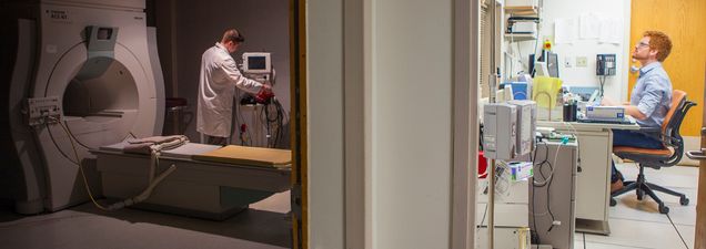
Students in the Bioimaging program benefit from hands-on experience with 1.5 and 3.0T MRI, which will ultimately qualify them for positions in the healthcare and biomedical instrumentation industries, image analysis, pre-clinical imaging, academia, and in a wide variety of private and government research centers. To satisfy the demand for the field’s broad exposure to imaging modalities, we provide students with a choice between two tracks to obtaining their degree.
Program Objectives and Outcomes
The goal of the program is to train professionals in all aspects of bioimaging from theory to practice. The field of bioimaging has become a major tool for the disciplines of clinical medicine, biomedical research, and image processing. The acceleration of imaging technology has been remarkable, particularly in areas of MRI, CT, and PET. Find out what some of our students are doing to learn about the career opportunities available to program graduates.
A Field in Constant Evolution
In the world of health delivery, the term MRI has become commonplace. It has entered our everyday language as more individuals find themselves getting an MRI for a variety of symptomatologies related to almost every organ in the body. Among conditions now routinely detected by MRI are:
- disorders of the brain, including tumors
- AV malformations
- aneurisms
- white matter lesions
- strokes
Studies of cardiac function and integrity are also now routinely performed with MRI and CT. In fact, several centers are now proactive in providing CT and MRI imaging to healthy individuals as a general screening device. MRI guided ultrasound is now used to identify, target and destroy circumscribed cancers in many body organs such as the liver.
With respect to research, imaging tools seem to be limitless in their application:
- Biomedical researchers are now viewing organs, tissue and body cavities at a level of detail approaching histologic quality
- With fMRI, brain activity can now be visualized over a wide range of cognitive and behavioral functions
- DTI provides our first glimpse into the neural connectivity of the human brain
- Cardiac MR and MR angiography provides a powerful tool for the study of heart function in health and disease
Thus, the demand for individuals skilled in the background, mechanics, operation, and interpretation of imaging techniques in a research or medical setting is growing at accelerated rates.![]()
Two Degree Options
Our Master of Science in Bioimaging program offers two 36 credit one-year degree options: our research path and our clinical path. Each path provides a broad background in basic and advanced sub-disciplines of imaging and anatomy, embodying the latest advances in imaging technologies, neurosciences, and robotics. The two one-year tracks differ in their goals for the preparation of individuals entering the field.
- The Clinical Path provides students with the didactic education and ethics requirements necessary to sit for the ARRT advanced certification exam in MRI. This certification allows an individual to enter the bioimaging field as a Registered MRI Technologist.
- The Research Path provides students with a research-based focus, culminating in a thesis project that preparing the individual for entry into the broader fields of academia, industry, image analysis, pre-clinical imaging, and beyond.