BU’s Cryogenic Electron Microscopy (CryoEM) Core Facility: Understanding the structures of life at a near-atomic scale
Studying the biological structures of viruses, proteins and other tiny macromolecules is key to understanding how life systems function and what happens when disease or injury cause these systems to break down.
These macromolecules are so small that traditional microscopes can’t see them very well. Instead, it takes a special kind of microscope to view these samples at the scale needed to fully understand them. The more powerful the microscope, the better researchers are able to map out the samples’ unique structures and applications.

Scientists at Boston University have the tools to do just that. Enter the Cryogenic Electron Microscopy (CryoEM) Core Facility, launched in April 2024 at the Center for Advanced Biomedical Research on the BU Medical Campus.
The cryoEM facility allows research teams from across BU to determine structures of macromolecules at near-atomic scale. By quickly freezing them in liquid nitrogen-cooled liquid ethane, the samples freeze as a glass in an almost native state.
“A car has all these parts that make it run properly,” said Professor of Pharmacology, Physiology & Biophysics Esther Bullitt, PhD, in a previous Spotlight interview. “It’s the same thing with bacteria, viruses and human beings.”
“To figure out how it works, we look at the pieces. If you can determine their structures at high enough resolution, then you can learn a lot about how it works,” Bullitt said.
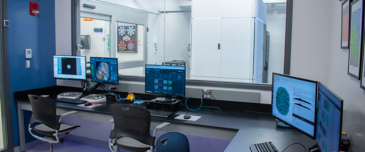
The cryoEM microscope collects hundreds of images per hour of each frozen sample, which are then analyzed and aligned into a 3D structure of each sample. Researchers can use these precise 3D structures to understand and advance their research in health and disease even further.
The facility was funded partially by a National Institutes of Health Shared Instrumentation Grant, awarded to Bullitt, who also serves as the Core Facility’s director. CryoEM Scientist Chad Hicks, PhD, serves as the Core Facility’s manager, and Professor of Pharmacology, Physiology & Biophysics Christopher W. Akey, PhD, serves as the facility’s advisor.
Learn more about our CryoEM Core Facility, and its applications to our researchers’ studies across BU, at the links below:
GMS Faculty Spotlight: Esther Bullitt, PhD
Dr. Bullitt is a professor of Pharmacology, Physiology & Biophysics at Boston University Chobanian & Avedisian School of Medicine and the director of both the biophysics and physiology graduate programs within Graduate Medical Sciences. She was awarded a Shared Instrumentation Grant by the National Institutes of Health, which helped to fund BU’s new CryoEM Core Facility.
Celebrating the “First Light” of the New CryoEM
A “First Light” gathering was held in April 2024 to celebrate the inaugural electron beam from the CryoEM. Learn more about the event here.
CryoEM of cardiac AL-224L amyloid reveals shared features in λ6 light chain fibril folds
BU scientists recently submitted a preprint of a high resolution cryoEM structure obtained at the CryoEM Core Facility. The structure is a light chain amyloid protein fibril obtained from a patient’s heart tissue. The work was a collaborative effort with BU’s Amyloidosis Center and helps us understand the deadly disease of Amyloidosis.
Boston University CryoEM Core Facility
Check out the CryoEM Core, along with details on its instruments, prices, policies, training and educational resources.