Researchers Develop Simple, Noninvasive Way to Assess Collagen in the Skin of Scleroderma Patients
Forearm skin biopsy sections stained with Masson’s trichrome, which highlights collagen in blue. Pathologists use these stains to assess skin fibrosis in scleroderma.
Convergent Research
Researchers Develop Simple, Noninvasive Way to Assess Collagen in the Skin of Scleroderma Patients
Earlier diagnosis and treatment help improve prognosis and survival.
Scleroderma (systemic sclerosis) is a rare, chronic, autoimmune disease that causes the body to produce too much collagen, leading to the hardening and thickening of the skin and internal organs. For more than 50 years, physicians have relied on manually pinching the skin to assess its thickness, a subjective approach that remains central to the diagnosis of systemic sclerosis, assessment of disease extent and progression, and monitoring of response to treatment. Despite being the accepted standard practice, doctors can score the same patient differently based on their own unique touch and judgement, making it difficult to accurately track changes in skin thickening over time or assess a patient’s response to treatment.
We have been developing SFDI for almost 15 years in my lab, but this is the first time anyone has applied this technology to scleroderma.
Darren Roblyer, PhD
For the first time, researchers at Boston University Chobanian & Avedisian School of Medicine and the Boston University Department of Biomedical Engineering have developed and tested a camera-based technique called spatial frequency domain imaging (SFDI) to assess skin changes in scleroderma patients. SFDI works by shining patterns of harmless light on the skin, without touching it, and measuring how the light bounces back. “We have been developing SFDI for almost 15 years in my lab, but this is the first time anyone has applied this technology to scleroderma” explained Darren Roblyer, PhD, professor of biomedical engineering and co-author on the manuscript.
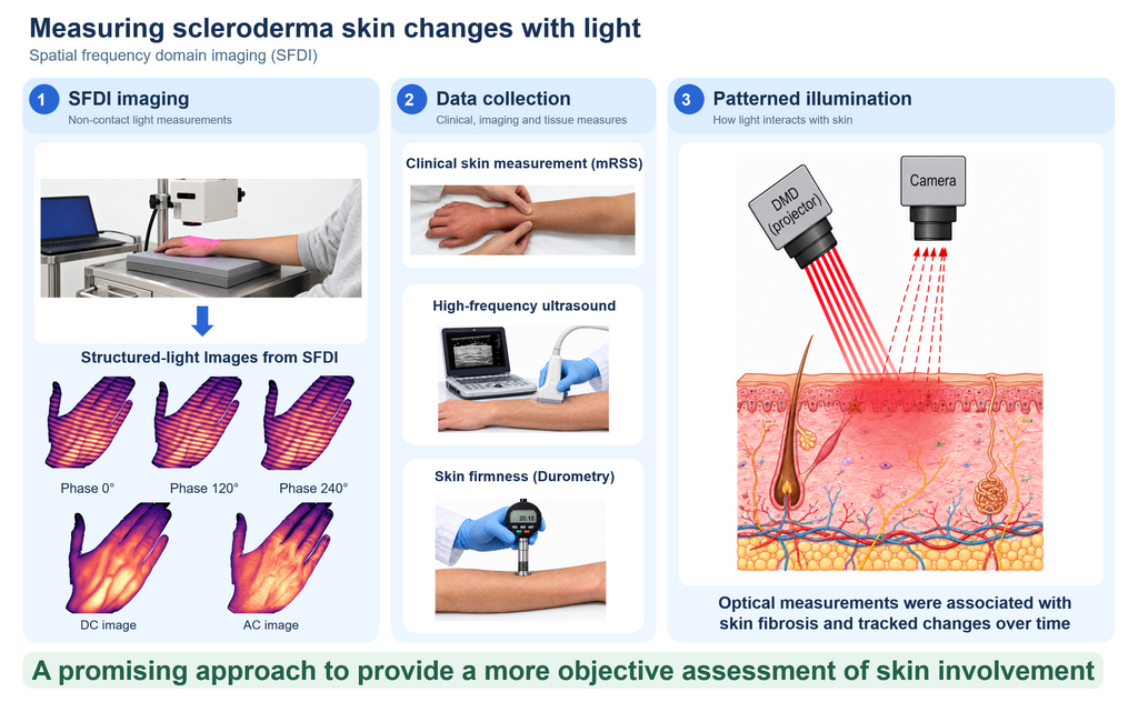
The researchers found that the light readings were different in people with scleroderma than in healthy individuals, and that they were able to track how thick and fibrotic (scarred) the skin was. They also found readings were different in skin that doctors had scored as normal, suggesting that SFDI may identify subtle skin changes not apparent on a standard clinical examination.
 Andreea Bujor, MD
“We compared the light readings directly with the amount of collagen buildup in skin biopsies, linking the imaging measurements to the scarring in the skin. We also compared the readings with ultrasound skin thickness and the standard clinical skin score, tested how repeatable the measurements were, and examined whether they changed as patients’ skin changed over time,” explained corresponding author Andreea Bujor, MD, associate professor of medicine at the school.
The researchers studied 49 people with scleroderma and 25 healthy volunteers at Boston Medical Center (BMC) Scleroderma Clinic, using SFDI to image six sites across the hands, forearms and fingers. They compared the light readings with the doctor’s touch-based skin score, ultrasound measurements of skin thickness, a handheld measure of skin hardness, and forearm biopsies from 34 patients and 16 healthy volunteers. In 11 patients, they repeated measurements over time to see whether the light readings tracked changes in the clinical skin score. Light readings were lower in people with scleroderma and lower readings were associated with higher skin scores, more scarring in biopsies, and thicker skin on ultrasound.
A fast, painless, non-contact measurement could help doctors track skin changes more objectively, help clinical trials detect treatment benefits more reliably and identify subtle skin changes before they can be felt by hand.
Andreea Bujor, MD
There are currently no approved therapies specifically for skin fibrosis in scleroderma. A major barrier to developing effective treatments is the lack of precise, objective methods to measure skin fibrosis, which can make even a beneficial therapy appear ineffective in clinical trials. “A fast, painless, non-contact measurement could help doctors track skin changes more objectively, help clinical trials detect treatment benefits more reliably and identify subtle skin changes before they can be felt by hand,” said Bujor, who also is a rheumatologist at BMC.
These findings appear online in the journal The Lancet Rheumatology.