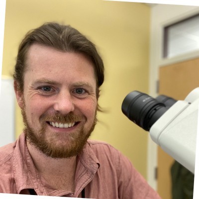
Nicholas Crossland
Assistant Professor
Assistant Professor of Pathology,Assistant Professor of Virology, Immunology & Microbiology
Link to BU Profile
Research Interest
In our lab, we focus on studying viral pathogenesis and evaluating novel medical countermeasures against emerging pathogens with significant public health implications. Our primary goal is to understand the intricate dynamics of viral-host pathogen interactions, with a special emphasis on identifying potential drug targets. We prioritize the comprehensive understanding of pathogenesis as the cornerstone of our research efforts.
Furthermore, we emphasize the critical need to rigorously validate pre-clinical models to ensure their fidelity in replicating human diseases. Our lab operates at the nexus of academia and industry, engaging in multi-disciplinary research endeavors aimed at addressing pressing infectious disease challenges.
Through the fusion of accumulated expertise, innovative technologies, and meticulous methodologies, we transcend conventional norms of a comparative pathology laboratory. Our approach enables us to delve deeply into the complexities of viral pathogenesis and evaluate the efficacy of medical countermeasures with unparalleled precision and depth.
Methodologies utilized by the NEIDL Comparative Pathology Laboratory
Routine Histology Processing of Formalin Fixed Paraffin Embedded (FFPE) Blocks: We employ a meticulously refined histology processing protocol, resulting in the generation of formalin-fixed paraffin-embedded blocks to unveil the intricate details embedded within.
Qualitative and Semi-Quantitative Histopathologic Analysis of All Organ Systems: Our seasoned team conducts exhaustive qualitative and semi-quantitative histopathologic analyses across diverse organ systems, meticulously scrutinizing every microscopic detail to unravel the nuances of pathological processes.
Cytochemical Stains, Automated High Plex Immunohistochemistry, and In Situ Hybridization: Leveraging classical approaches with the latest technological advancements, we harness cytochemical stains, automate high plex immunohistochemistry, and in situ hybridization techniques to delve deep into molecular signatures, unraveling the intricate tapestry of pathogen-host cellular interactions.
Multispectral Whole Slide Scanning and Quantitative Image Analysis: Through state-of-the-art multispectral whole slide scanning technology, we capture comprehensive images, allowing for nuanced analysis and precise quantification. Our quantitative image analysis techniques unravel layers of complexity, unveiling insights inaccessible through conventional means.
Spatial Transcriptomics (10x Visium Platform): At the forefront of innovation, we integrate spatial transcriptomics using the groundbreaking 10x Visium platform. This enables us to decode the spatial organization of gene expression within tissue microenvironments, illuminating previously obscured patterns and interactions.