News
Resident Perspective
Dr. Osamu Sakai, Dr. Carlota Andreu-Arasa, Dr. Baojun Li and Resident Dr. Pedro Staziaki Published in the European Journal of Radiology
[av_textblock size='' av-medium-font-size='' av-small-font-size='' av-mini-font-size='' font_color='' color='' id='' custom_class='' av_uid='av-kd7qmyzm' admin_preview_bg='']
Using CT texture analysis to differentiate cystic and cystic-appearing odontogenic lesions
Abstract
Purpose: Cystic and cystic-appearing odontogenic lesions of the jaw may appear similar on CT imaging. Accurate diagnosis is of

ten difficult although the relationship of the lesion to the tooth root or crown may offer a clue to the etiology. The purpose of this study was to evaluate CT texture analysis as an aid in differentiating cystic and cystic-appearing odontogenic lesions of the jaw.
Methods: This was an IRB-approved retrospective study including 42 pathology-proven dentigerous cysts, 37 odontogenic keratocysts, and 19 ameloblastomas. Each lesion was manually segmented on axial CT images, and textural features were analyzed using an in-house-developed Matlab-based texture analysis program that extracted 47 texture features from each segmented volume. Statistical analysis was performed comparing all pairs of the three types of lesions.
Results: Pairwise analysis revealed that nine histogram features, one GLCM feature, three GLRL features, two Laws features, four GLGM features and two Chi-square features showed significant differences between dentigerous cysts and odontogenic keratocysts. Four histogram features and one Chi-square feature showed significant differences between odontogenic keratocysts and ameloblastomas. Two histogram features showed significant differences between dentigerous cysts and ameloblastomas.
Conclusions: CT texture analysis may be useful as a noninvasive method to obtain additional quantitative information to differentiate cystic and cystic-appearing odontogenic lesions of the jaw.
Keywords
Ameloblastomas; CT; Dentigerous cysts; Jaw lesions; Odontogenic keratocysts; Texture analysis.
PMID: 31539792
DOI: 10.1016/j.ejrad.2019.108654
[/av_textblock]
Dr. Osamu Sakai, Dr. Carlota Andreu-Arasa, Dr. Baojun Li and Resident Dr. Pedro Staziaki Published in the European Journal of Radiology
[av_textblock size='' av-medium-font-size='' av-small-font-size='' av-mini-font-size='' font_color='' color='' id='' custom_class='' av_uid='av-kd7qmyzm' admin_preview_bg='']
Using CT texture analysis to differentiate cystic and cystic-appearing odontogenic lesions
Abstract
Purpose: Cystic and cystic-appearing odontogenic lesions of the jaw may appear similar on CT imaging. Accurate diagnosis is of

ten difficult although the relationship of the lesion to the tooth root or crown may offer a clue to the etiology. The purpose of this study was to evaluate CT texture analysis as an aid in differentiating cystic and cystic-appearing odontogenic lesions of the jaw.
Methods: This was an IRB-approved retrospective study including 42 pathology-proven dentigerous cysts, 37 odontogenic keratocysts, and 19 ameloblastomas. Each lesion was manually segmented on axial CT images, and textural features were analyzed using an in-house-developed Matlab-based texture analysis program that extracted 47 texture features from each segmented volume. Statistical analysis was performed comparing all pairs of the three types of lesions.
Results: Pairwise analysis revealed that nine histogram features, one GLCM feature, three GLRL features, two Laws features, four GLGM features and two Chi-square features showed significant differences between dentigerous cysts and odontogenic keratocysts. Four histogram features and one Chi-square feature showed significant differences between odontogenic keratocysts and ameloblastomas. Two histogram features showed significant differences between dentigerous cysts and ameloblastomas.
Conclusions: CT texture analysis may be useful as a noninvasive method to obtain additional quantitative information to differentiate cystic and cystic-appearing odontogenic lesions of the jaw.
Keywords
Ameloblastomas; CT; Dentigerous cysts; Jaw lesions; Odontogenic keratocysts; Texture analysis.
PMID: 31539792
DOI: 10.1016/j.ejrad.2019.108654
[/av_textblock]
Dr. Mohamad Abdalkader, Dr. Osamu Sakai and Dr. Thanh Nguyen Published in Interventional Neuroradiology
[av_textblock size='' av-medium-font-size='' av-small-font-size='' av-mini-font-size='' font_color='' color='' id='' custom_class='' av_uid='av-kd7qcg0n' admin_preview_bg='']
Endovascular coiling of large mastoid emissary vein causing pulsatile tinnitus
Abstract
The association of large mastoid emissary veins and pulsatile tinnitus has been reported. However, therapeutic options for this condition remain limited. We report a case of endovascular coiling of a large mastoid emissary vein in a patient with disabling pulsatile tinnitus with significant improvement of symptoms. To our knowledge, endovascular coiling of large mastoid emissary vein causing pulsatile tinnitus has not been reported.
Keywords
Pulsatile tinnitus; coil embolization; mastoid emissary vein.
PMID: 32408784
DOI: 10.1177/1591019920926333
[/av_textblock]
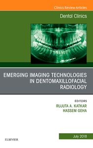
Dr. Osamu Sakai published in Emerging Imaging Technologies in Dentomaxillofacial Radiology

Multidector Row computed tomography in Maxillofacial Imaging
Gohel A1, Oda M2, Katkar AS3, Sakai O4.
Abstract
Multidetector row CT (MDCT) offers superior soft tissue characterization and is useful for diagnosis of odontogenic and nonodontogenic cysts and tumors, fibro-osseous lesions, inflammatory, malignancy, metastatic lesions, developmental abnormalities, and maxillofacial trauma. The rapid advances in MDCT technology, including perfusion CT, dual-energy CT, and texture analysis, will be an integrated anatomic and functional high-resolution scan, which will help in diagnosis of maxillofacial lesions and overall patient care.
Keywords
Dual-energy CT; Imaging; Maxillofacial; Multidetector row CT; Perfusion CT; Texture analysis
PMID: 29903561
Carlos Arellano recognized by BMC in this year’s ‘Be Exceptional Awards’
 Carlos Arellano, Senior Administrative Director of Otolaryngology and Radiology, is a recipient of the BMC’s Be Exceptional Award in Growth. Winners of this award demonstrate exceptional performance and dedication to fulfilling BMC’s mission and QUEST goals. These individuals also demonstrate initiative, teamwork, competence and embody BMC’s values of RESPECT.
Ali Guermazi Receives Clinical Research Award from Osteoarthritis Research Society International
Ali Guermazi, MD, PhD, professor and vice chairman of academic affairs is the recipient of Osteoarthritis Research Society International (OARSI) Clinical Research Award for his outstanding work in the field of imaging.
He is the first radiologist to receive this honor. His research interests include musculoskeletal diseases and in particular identifying structural risk factors for developing and worsening osteoarthritis.
The award was presented during the Opening Ceremony at the OARSI World Congress in Liverpool, England, on Friday, April 27. The OARSI Clinical Research Award recognizes excellence in clinical research related to osteoarthritis in areas such as epidemiology and health services research, clinical outcomes, translational research and therapeutics, encompassing all phases of research having clinical implications. OARSI is the premier international organization for scientists and health care professionals focused on the prevention and treatment of osteoarthritis through the promotion and presentation of research, education and the worldwide dissemination of new knowledge.
Discussions on the Opioid Epidemic
Dr. David Bates is interviewed by RadioGraphics editor Dr. Jeffrey Klein on how Radiology addresses the issue of the opioid epidemic. The interview references the article published in RadioGraphics titled “Acute Radiologic Manifestations of America’s Opioid Epidemic” which was co-authored by several BU Radiologists.
Dr. Osamu Sakai published in the journal RadioGraphics
The article titled “Craniofacial Manifestations of Systemic Disorders: CT and MR Imaging Findings and Imaging Approach” discusses imaging approaches for early detection of various systemic diseases or conditions that affect the maxillofacial bones.
