Trainee, Visitor and Volunteer Onboarding
CBM-led Study on Diagnosing Different Forms of Dementia Using Artificial Intelligence
(Boston)—Ten million new cases of dementia are diagnosed each year but the presence of different dementia forms and overlapping symptoms can complicate diagnosis and delivery of effective treatments. Now researchers from Boston University have developed an AI tool that can diagnose ten different types of dementia such as vascular dementia, Lewy body dementia,
and frontotemporal dementia, even if they co-occur.
The researchers created a multimodal Machine Learning (ML) framework that accurately identifies specific pathologies causing dementia using commonly collected clinical data, such as demographic information, patient- and family-level medical history, medication use, neurological and neuropsychological exam scores and neuroimaging data such as MRI scans. These findings appear online in Nature Medicine.
“Our generative AI tool enables differential dementia diagnosis using routinely collected clinical data, showing its potential as a scalable diagnostic tool for AD and related dementias,” says corresponding author Vijaya B. Kolachalama, PhD, FAHA, associate professor of medicine at Boston University Chobanian & Avedisian School of Medicine. “The ability to generate diagnosis with routine clinical data is becoming increasingly important given the significant
challenges in accessing gold-standard testing, not only in remote and economically developing regions and in urban healthcare centers,” adds Kolachalama who also is an associate professor of computer science, affiliate faculty of Hariri Institute for Computing, and a founding member of
the Faculty of Computing & Data Sciences at Boston University.
In the study, the multimodal ML framework was trained on data from more than 50,000 individuals from nine different global datasets. The model achieved an area under the receiver operating characteristic (ROC) curve of 0.96 in differentiating the dementia types. The ROC score can range from 0 to 1. A score of 0.5 indicates random guessing, and a score of 1
indicates perfect performance.
The team also compared the performance of neurologists and neuro-radiologists working alone versus with the AI tool and found that AI can boost the accuracy of neurologists by more than 26% across all 10 dementia types. Using 100 randomly selected cases, 12 neurologists were asked to make a diagnosis and provide a confidence score between 0 to 100. This confidence score was then averaged with the probability score obtained by the AI tool to obtain an AI augmented neurologist score.
“There aren’t enough neurology experts around the world, and the number of patients needing their help is growing quickly. This mismatch is putting a big strain on the healthcare system. We believe AI can help by identifying these disorders early and assisting doctors in managing their patients more effectively, preventing the diseases from getting worse,” adds Kolachalama.
With dementia cases set to double in the next 20 years, the researchers hope that this AI tool can provide accurate differential diagnosis and support the increased demand in targeted therapeutic interventions for dementia.
![]()
![]()
CBM Researchers to Co-lead Project to Intercept Lung Cancer
Lung cancer remains the leading cause of cancer deaths in the United States, with someone diagnosed approximately every two minutes. In a collaborative effort to end this devastating disease, the American Lung Association and LUNGevity Foundation have joined forces to invest $3 million over the next three years in research aimed at intercepting lung cancer – catching precancerous cells and blocking them from turning into cancer cells.

The partnership will fund research co-led by Ehab Billatos, MD, Jennifer Beane, PhD, Josh Campbell, PhD, Steven Dubinett, MD (UCLA), Sarah Mazzilli, PhD, and Avrum Spira, MD, MSc, under the project “Intercept Lung Cancer Through Immune, Imaging & Molecular Evaluation InTIME”. This research aims to establish a timeline of precancerous cell evolution into malignant cancer, utilizing cutting-edge technologies such as robot-assisted bronchoscopy to collect longitudinal samples from patients suspected to have lung cancer. Spira, Professor of Medicine and Global Head of Interventional Oncology at Johnson & Johnson, and Dubinett, Dean of the David Geffen School of Medicine at UCLA, are leaders in the field of lung cancer research.
This ongoing partnership brings together leading scientists dedicated to investigating early molecular and other changes that lead to cancer development, with the ultimate goal of stopping lung cancer before it begins. By focusing on early detection and intervention, the research aims to significantly improve survival rates and outcomes for patients.
“Early detection of lung cancer is crucial to saving lives. By investing in research to intercept lung cancer at its earliest stages, we have the potential to revolutionize how we approach this disease and improve outcomes for patients,” said Harold Wimmer, president and CEO of the American Lung Association.
“Continuing our partnership with the American Lung Association to help stage-shift lung cancer underscores our commitment to advancing research that will make a tangible difference in the lives of those affected by the disease. Together, we are harnessing the power of scientific innovation to drive progress in interception strategies and potentially cure lung cancer,” said Andrea Ferris, president and CEO of LUNGevity Foundation.
“This project represents an evolution of our ongoing efforts to understand and intercept lung cancer before it progresses,” said Spira. “By unraveling the molecular and immune mechanisms underlying lung cancer development, we can develop targeted strategies for early detection and intervention.”
The partnership builds upon previous collaborations, including research funded by the American Lung Association, LUNGevity Foundation, and Stand Up to Cancer, which has yielded significant findings in lung cancer interception. Notable achievements include mapping the precancer genome, the discovery of enzymes and proteins associated with premalignant cells, and offering potential targets for treatment, as well as insights into immune cell activity in premalignant lesions. Partial funding was provided by the Thomas G. Labreque Foundation.
Through initiatives like the American Lung Association Research Institute Accelerator Program and the LUNGevity Early Lung Cancer Center, this partnership aims to accelerate progress in lung cancer interception research and pave the way for personalized treatments for patients.
Maria Antoinette Evans Staff Diversity Award 2023-24
Erin Kane, PhD, Scientific Program Manager has been awarded the Department of Medicine’s Maria Antoinette Evans Staff Diversity Award which was created to honor a non-faculty member who promotes the departmental values of diversity, equity, inclusion, and accessibility in the DOM or in the broader community. Erin is the chair of the section’s DEIA Committee, which includes a cross-section of faculty, staff, and trainees. The committee’s goal is to communicate and organize DEIA opportunities to support the well-being of all section members.
Congratulations to Erin on winning this year’s award!
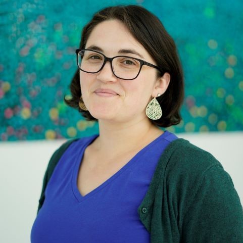
Researchers Shed Light on Signaling Pathway Responsible for Head and Neck Cancers
A new study from researchers at Boston University Chobanian & Avedisian School of Medicine shows how computational approaches can drive important biomedical discoveries. The study aimed to better characterize oral tumor heterogeneity, including aggressive cell subpopulations, and identify candidate vulnerabilities that could be targeted therapeutically. The findings are of timely significance, providing new information about additional therapeutic targets for head and neck cancer. The findings appear online in the journal Translational Research.
To read the press release, click here.
Centenarians Possess Unique Immunity that Helps them Achieve Exceptional Longevity
Led by researchers from Boston University Chobanian & Avedisian School of Medicine and Tufts Medical Center, a new study finds centenarians harbor distinct immune cell type composition and activity and possess highly functional immune systems that have successfully adapted to a history of sickness allowing for exceptional longevity. These immune cells may help identify important mechanisms to recover from disease and promote longevity. The findings appear online in the journal Lancet eBiomedicine.
New Hires May 2023
Grace Kennedy is a summer Research Study Assistant working with the clinical research team. She is an undergraduate student at the University of Massachusetts Lowell and expects to graduate with a Bachelor of Science in Biological Sciences in 2025.

Marissa Flynn has also joined the clinical research team as a Research Study Assistant. She graduated with a Bachelor of Arts degree from the University of Vermont in May 2020. Marissa gained research experience working at the University of Vermont Medical Center during her undergraduate program. She has relocated to Boston from Lee, New Hampshire.

Jamali is a new Postdoctoral Associate working with Ignaty Leshchiner. He received his Ph.D. in Statistical Physics from Shahid Beheshti University in 2015. Tayeb has joined us from Kansas State University where he was a postdoctoral research associate. Anqi will be working on K4.

Anqi Zou joined Chao Zhang’s lab as a Postdoctoral Associate on May 22. She received her PhD in Solid Earth Physics from the University of Chinese Academy of Sciences, Beijing, China in 2022 and was most recently a research engineer at the Institute of Geology and Geophysics, Chinese Academy of Sciences. Anqi will be working on K4 with Chao and his research team
Moorman-Simon Fellowship 2022-2023
The Moorman-Simon Fellowship in Computational Biomedicine was established in the summer of 2019 to provide fellowships to current Ph.D. students in Computational Biomedicine who are working in the field of cancer research. This fund was established through a generous gift by Trustee Ruth A. Moorman (CAS’88, Wheelock’89,’09, Parent BUA’15) and Sheldon N. Simon.
Congratulations to Conor Shea and Charley Xi, the recipients of the 2022-2023 Moorman Simon Fellowship. Please see below for more information on their projects.
Conor Shea, MD/PhD Candidate, Beane and Spira-Lenburg Labs

Lung cancer is the most common cause of cancer death in the United States and the world. Squamous cell carcinoma, the second most common subtype of lung cancer, is preceded by precursors ("premalignant lesions", "PMLs"), which could be targeted to intercept lung cancer and improve patient mortality. While we know that most of these PMLs will not progress to cancer, we are currently unable to a) predict which patients' PMLs will progress to cancer, and b) intercept PMLs before they become cancer. Our lack of knowledge about the biology of PMLs limits our ability to accomplish these goals, and my project seeks to address those knowledge gaps. In my thesis work, I use single cell RNA transcriptomics to profile the PML cellular ecosystem and identify changes associated with each step ("grade") of PML cancerization. Already, I have identified PML grade-associated changes in several cell types. I am also combining my single cell RNA transcriptomics data with previous work in the lab to study gene expression signatures associated with PML expression. The findings in this project can be used to improve lung cancer mortality by identifying patients at highest risk of cancer and mechanisms for PML interception.
Zhan (Charley) Xi, PhD Candidate, Beane and Mazzilli Labs
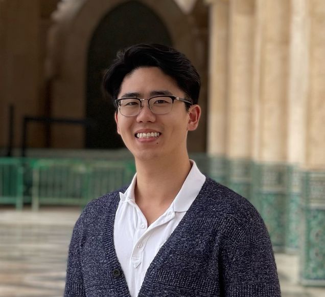
The goal of my project is to study the tumor immune microenvironment (TIME) of lung cancer, which may be used to develop enhanced approaches to assess the progression and severity of early lung cancer and to suggest early intervention strategies. To do so, I am using single cell RNA sequencing (scRNA-seq) and Cellular Indexing of Transcriptomes and Epitopes by Sequencing (CITE-Seq) to study the molecular changes at a single cell level, including the changes in protein and gene expression that dictate a cell’s activity. Then I will use imaging mass cytometry (IMC) to identify the spatial locations of the cells identified within the lung cancer tissue and use computational methods to decipher the interactions among the different types of immune cells, and how they collaborate as a group to suppress or enhance the tumor’s progression. Ultimately, our goal for future patients is being able to accurately diagnose the severity and progression of lung cancer to reduce cancer mortality.
Two Technologies That Can Make Diagnosing Dementia Easier for Doctors and Patients
(Source: The Brink)
Dr. Vijaya Kolachalama, Associate Professor of Medicine and CBM faculty member, was quoted in the Brink article "Two Technologies That Can Make Diagnosing Dementia Easier for Doctors and Patients." The article also featured more information about a deep learning algorithm he and his team developed. See below for a an article preview and click the link to read the full article.
Predicting Alzheimer’s with Artificial Intelligence
To diagnose dementia, physicians typically perform cognitive tests on attention, memory, problem-solving, and other abilities, along with a physical exam, blood tests, and brain scans. This battery of evaluations can help determine whether the memory issues a person is experiencing are caused by Alzheimer’s or another form of dementia.
“Patients walk in and doctors have to try to understand where they fall on the dementia spectrum,” says Vijaya B. Kolachalama, a BU School of Medicine assistant professor and expert on using computers to aid medical diagnoses. He and his team developed a deep learning algorithm that, for the first time, attempts to predict where a person falls on the dementia spectrum and identify if their memory loss is due to dementia or other reasons.
New Publication from Campbell Lab
(source: Nature Communications)
The Campbell lab, led by its Bioinformatics students, has just been published for SCTK-QC, a pipeline for comprehensive QC of single-cell RNA-seq data. This pipeline will help with reprocessing and QC efforts in consortiums. Click here to read the full publication.
Identifying New Biomarkers to Detect Lung Cancer Earlier
Source: NIH National Cancer Institute, Division of Cancer Prevention
Lung cancer, the leading cause of cancer deaths worldwide killing 1.8 million people each year, is often diagnosed at an advanced stage when the chances for a cure are limited.
In the United States, almost 60% of people diagnosed with localized lung and bronchus cancer are likely to survive for 5 years. This is nearly 10 times more than those who are not diagnosed until their cancer has spread to distant parts of the body, who have a 5-year relative survival of 6.3%. Right now, more than half of lung cancers are diagnosed at this late state.
Researchers are working to change that with a precision approach to early detection.
“Detecting lung early is very important for decreasing mortality,” said Jennifer Beane, Ph.D., a computational biologist at Boston University School of Medicine. “If you detect lung cancers early, you can resect them for cure.”
The U.S. Preventive Services Task Force recommends annual screening by low-dose computed tomography (CT) for adults aged 50 to 80 years with a 20 pack-year smoking history (averaging a pack of cigarettes a day for 20 years) who currently smoke or have quit within the past 15 years. But these criteria don’t cover everyone who develops lung cancer.
“Only 25% to 30% of patients who have lung cancer would have been captured by the current lung cancer screening guidelines,” said Ehab Billatos, M.D., a pulmonologist at Boston University School of Medicine. “We're missing a lot of patients.”
Click here to read more.