Gross Anatomy Lab Renovations Looking Good
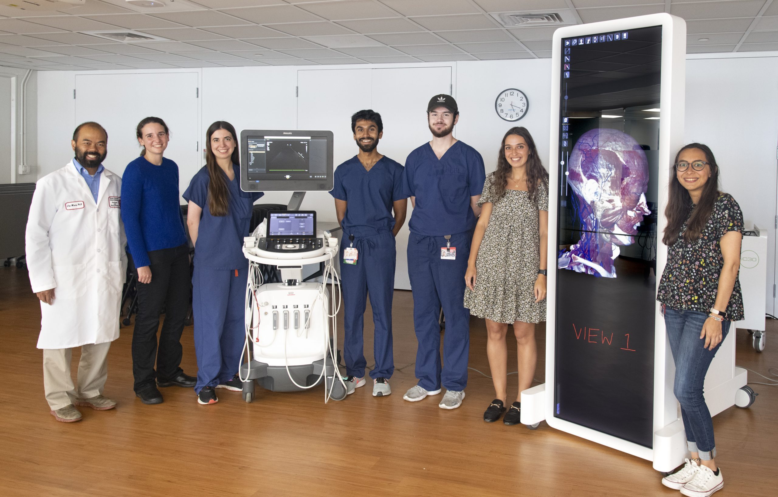 (L-R) On a recent tour of the renovated Gross Anatomy Lab, Jonathan Wisco, PhD (BUSM’02), associate professor of anatomy & neurobiology, medical students Jessica Landau-Taylor, Lindsey Claus, and Nimish Saxena and teaching assistants Tyler Capen, Sydney Mosaheb, and Paola Castro stand with a new ultrasound machine and a new Anatomage virtual dissection table.
There’s a process to becoming a doctor and anatomy class is one of the steps to getting there. First-year medical school students face a tsunami of names and functions for a dizzying array of body parts that they must learn and understand before proceeding on to their clinical education where they see patients.
“If we taught you how to ride a bicycle the way we’ve traditionally taught anatomy, you’d have to learn every part of the bicycle before you actually got on it,” said Jonathan Wisco, PhD (BUSM ’02), associate professor of anatomy & neurobiology.
Thanks to a significant gift from donors who wish to remain anonymous, a $1 million overhaul of the Gross Anatomy Lab began in 2020 and is nearly complete. Wisco said the anatomy lab upgrade recognizes that there are multiple pathways to learning and was in step with the new curriculum changes implemented this year for the Class of 2026 that incorporate self-directed learning and early exposure to clinical concepts.
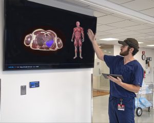 Tyler Capen, a Gross Anatomy Lab research assistant and second-year anatomy and neurobiology graduate student, demonstrates the new VH Dissector that displays 3D and cross-sectional views of over 2,000 anatomical structures.
“What we’re doing is we’re providing students the exposure to the 3D donor, plus the technology, before they dissect,” said Wisco. “The more that students know what they are looking for, the better dissectors they are.”
New windows flood the 10th-floor anatomy lab with sunlight, including the portion where dozens of donor bodies lie in bright blue body bags on portable dissection tables.
“The natural sunlight coming in puts your mind at ease,” said Anatomy Laboratory Director and Anatomical Gift Director Robert Bouchie. “It shouldn’t be macabre; it should be something that you’re embracing.”
As many as 45 donor bodies are used every year for between 309 and 370 students, with as many as 180 medical students and 130 dental students participating. When the lab is in session it’s noisy, said Bouchie, but it’s a good noise – the sound of students learning how they can help future patients.
“I want there to always be this subliminal feeling of life,” he said.
The upgrades include improved communications for instructors with an enhanced sound system and monitors linked to iPads, as well as updated lighting, heating, air conditioning and ventilation. New technology includes ultrasound units and an Anatomage virtual dissection table. The communications upgrades allowed images from the VH Dissector, that displays 3D and cross-sectional views of more than 2,000 anatomical structures, to be displayed on monitors across the lab.
Wisco encourages students to use both the traditional teaching aids like anatomy books and photos and new technologies to prepare for a dissection on donor bodies and to work backwards from the dissection through the technology to textbooks to better understand what they encountered.
“That way they are contextualizing more and more sophisticated information that they imprint in their mind,” said Wisco.
With new technologies that can duplicate dissection, third-year medical student Jessica Landau-Taylor was initially skeptical about the need for real donor bodies. But she found the new technology in the lab worked well with dissection, improving the process by providing context and the opportunity for self-directed learning.
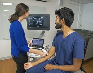 Third-year medical student Jessica Landau-Taylor uses a new ultrasound machine on second-year medical student Nimish Saxena in the newly-renovated Gross Anatomy Lab. Students use the ultrasound to see how organs and other bodily systems look and work in a living person.
“There’s nothing quite like actually being able to see the structure (tissues, organs etc.) in an actual human being, in color, with your eyes, the same way you see everything else,” said Landau-Taylor. “It’s also good to see it in a way that’s been formatted to help you learn.”
“(The Anatomage table) is a great way to visualize anatomy, especially anatomy that may be difficult to see on a cadaver,” said post-graduate teaching assistant Sydney Mosaheb, MS, who also is a research assistant in the CTE Center. The technology makes it easy for students to investigate on their own or double-check their work on the human donor.
“Trial and error, at least for me, is helpful for learning and they (medical students) have to learn so many things that being able to just memorize it in a way that makes sense to them is very helpful,” said teaching assistant Paola Castro, MS, who also is a research technician in the Cellular Neurobiology Lab.
The new technologies also lend color and a realistic look to organs and other structures that can be relatively colorless and physically flattened in donor bodies due to the embalming process. The ultrasound, VH Dissector and the Anatomage also help students interpret the images from similar instruments like ultrasound, Magnetic Resonance Imaging (MRI) and Computed Tomography (CT) scans students will see in their clinical rotations and as physicians.
“I had no radiology experience, and this (VH Dissector) made it click for me when I first saw an MRI,” said teaching assistant and second-year anatomy & neurobiology graduate student Tyler Capen. “An MRI is a lot of black and white but if you know what the structures look like, what shapes they are and where they’re supposed to be, you can correlate that.”
Second-year medical student Nimish Saxena found the technology helped inject excitement into the daily routine of lectures and rote memorization. Employing technology that resembles what they will use as clinicians gave the anatomy courses more relevance, he said.
“It’s really good for melding that clinical relevance with the coursework we’re studying,” said Saxena.
View all posts