Gross Anatomy Lab Gets Facelift
Thanks to a generous gift by Albert and Debbie Rosenthaler, BUSM’s Gross Anatomy Lab is getting a major facelift.
According to Waterhouse Professor and Anatomy & Neurobiology Chair Jennifer I. Luebke, PhD, this financial gift allowed the department to make needed renovations to the lab’s infrastructure. A new heating, ventilation and air conditioning (HVAC) system is being installed to enhance air quality and reduce noise. Visibility in the lab also is being improved with the addition of bright LED lights in a sleek new suspended ceiling.
The donation also helps the lab to go a step further and attain the most up-to-date teaching tools. “Taken together, the enhancements to the Gross Anatomy Lab made possible by this very generous gift will greatly facilitate medical education at the School of Medicine across all four years of education,” said Dr. Luebke.
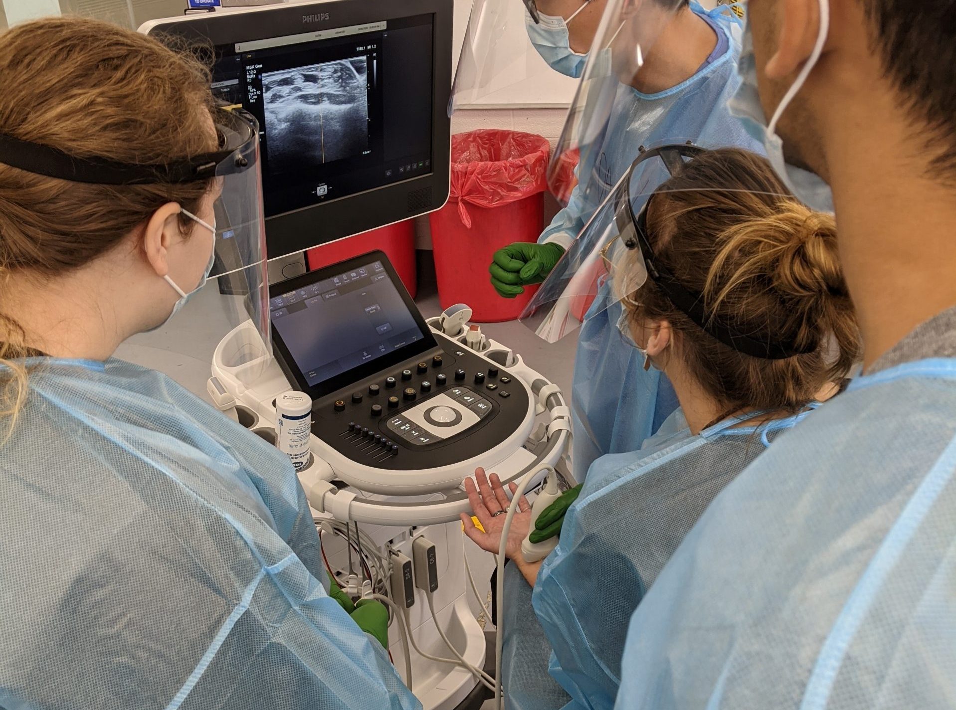
 A fourth-year student ultrasounds the wrist of a first-year student during anatomy lab.
One of the new modern teaching tools is a state-of-the-art Ultrasound System, which enables teaching anatomy and clinical cases on both live subjects and cadavers. This sophisticated imaging modality is commonly used in Emergency Medicine as a quick, non-invasive way to image structures close to the skin. Ultrasound imaging is helpful in primary care settings as well, and has historically been used in obstetrics and gynecology.
“It is very exciting to have ultrasounds in the anatomy lab, because it allows us to incorporate clinical imaging modalities into the students’ education as soon as medical school begins,” said Associate Professor of Anatomy & Neurobiology Ann Zumwalt, PhD. “Thanks to the generosity of the donor, we were able to get three full-sized units, which will allow the students to interact with units that are essentially identical to what they will see in the clinic.”
This donation also has permitted the lab to install monitors and cameras, making it possible for the entire class to visualize the anatomical structures in a given cadaver. Faculty now will be able to effectively communicate visual material to the entire class simultaneously.
“From an anatomy education perspective, it is exciting to be able to teach about anatomical structures and then immediately image them on either living subjects or on the anatomical donors,” said Dr. Zumwalt.
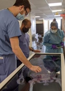
 Dr, John Wisco and 4th year student instructors Ben Weaver and Priyanka Jethwani explore the features of the table.
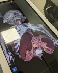
 The Anatomage table enables 3D reconstruction of cadaveric anatomy in real life size. During the week the class was studying the abdomen, the table was used to simultaneously examine three-dimensional and cross-sectional anatomy.
Last but not least, a new virtual dissection table will give students the ability to access views of the human body that are impossible with the cadaver. Users are able to visualize real human anatomy without the chemicals and smells from a traditional cadaver. As noted by Anatomage, the company that produces these tables, it is “the most technologically advanced 3D visualization system for anatomy and physiology education and is being adopted by many of the world’s leading medical schools and institutions.”
“This acquisition aligns us with the trajectory of where anatomy education is heading nationally,” said Dr. Zumwalt. “I’m really excited about the opportunities this opens up for us!”
Submitted by Sara Frazier.
View all posts