PhenoImager HT Quantitative Pathology Imaging System
The PhenoImager HT is an upgraded version of the Vectra Polaris image analysis suite, capable of unmixing whole slide images (WSI) during the scanning process. This enables users to perform quantitative analysis such as positive pixel analysis, cell phenotyping, and spatial analysis.
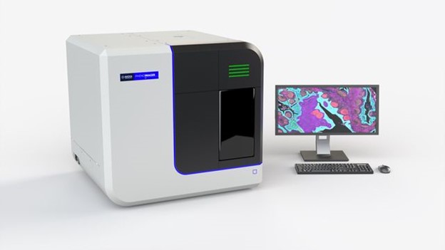 Akoya Biosciences PhenoImager HT Whole Slide Imaging System
Overall Benefit
The addition of the PhenoImager HT Quantitative Pathology Imaging System to the BU medical campus will enhance research capabilities by:
- High-Throughput Imaging: This system allows for rapid whole slide image acquisition of biological specimens, capturing both brightfield and high-plex fluorescent images (up to six colors plus DAPI). This capability enables more efficient utilization of existing research samples.
- Improved Analytical Sensitivity: By employing multispectral unmixing, the system increases signal-to-noise ratios and effectively removes autofluorescence signals, leading to clearer and more reliable imaging results.
- Quantitative Analysis: The integrated image acquisition and advanced analysis pipeline provide precise and objective characterization of morphomolecular signatures, placing them in spatial context for a deeper understanding of biological processes.
- Comprehensive Pathomics Datasets: The system generates continuous pathomics datasets that can be correlated with other biological data (e.g., clinicopathologic, transcriptomic). This integration facilitates more rigorous research approaches, helping to clarify the clinicopathologic correlates of diseases.
Akoya Biosciences PhenoImager HT Specifications
PhenoImager HT Epifluorescence Filter Cube Characteristics
Recommended Fluorescent Dyes
Services provided by staff include:
- Core competency training on the operation of the PhenoImager HT image acquisition.
- Assisted or independent use of the PhenoImager HT whole slide scanner, with the latter only available to those approved by staff.
- Guidance in generating multispectral libraries to remove auto-fluorescence and maximize signal to noise ratios of images using inForm.
- Assistance and training in the generation of quantitative pathology outputs using inForm.
Services
|
BU Internal Rates |
External Rates |
| Brightfield Unassisted/Slide |
$16 |
$28 |
| Brightfield Assisted/Slide |
$20 |
$36 |
| Fluorescence Unassisted/Slide |
$30 |
$64 |
| Fluorescence Assisted/Slide |
$36 |
$54 |
How to Schedule
Please login to iLab Scheduling System to schedule equipment time or services. For new users please follow the steps outlined in Information for New Users.
Usage and Training
Training is mandatory for all new users and requires:
- Scheduling of two consecutive 1-hour sessions of “Assisted Use” for practical in-person training.
- Users are expected to bring their own slides for scanning during the training session.
- A combination of both brightfield and fluorescent slides is recommended to familiarize users with developing scan profiles for each of these formats.
Please contact us to schedule training or assisted use. Once users have been trained, time on the instruments may be scheduled.
We require that all users cancel sessions 24 hours before the scheduled start time. Failure to cancel will result in the user (or PI) being billed for the entire scheduled duration.
Acknowledgments
The PhenoImager HT was funded by the NIH Award Number S10OD030269. We greatly appreciate acknowledgement of NIH S10OD030269 in publications when authors use our equipment and/or assistance in their research.
Contact
Nicholas Crossland, DVM, DACVP
Director
(617) 358-9285 | ncrossla@bu.edu
Hans Gertje, BS, HTL(ASCP)CMQIHCCM
Lab Supervisor
(617) 358-9139 | hgertje@bu.edu
Location
Boston University Chobanian & Avedisian School of Medicine
Housman Medical Research Center
72 East Concord Street, R Building, R-824A
Boston, MA 02118
View BU Medical Campus MAPS
◄ Back to Integrated Biomedical Imaging Services (IBIS) website
BACK TO TOP↑