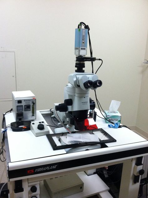
Olympus Stereo Fluorescence Imaging Microscope

Function of the instrument
This system is a low magnification white light and fluorescence upright imaging microscope suitable for imaging embryos, organs and tissue samples.
Overview
This system is a low magnification stereo microscope ideal for imaging live embryos and whole organs. It has a computer controlled motorized z-axis drive for the automated acquisition of images in multiple planes. It uses a mercury-halide fluorescence light source and filters for fluorescence imaging of nuclear stains as well as expressed fluorescent proteins. The system is equipped with a cooled monochrome CCD camera for high-sensitivity fluorescence imaging. The camera can also be used for transmitted light and oblique illumination images. Color images can be obtained using an RGB LCD electronic emission filter. A microincubation system allows for time-lapse embryo or organ culture experiments.
- Training involves proper turn on and turn off sequences.
- It also involves learning the proper operation and cleaning so as not to damage the optics or other parts of the instrument.
- Generally, the instructor is operating the instrument for the first session and explaining as the experiment progresses and is present for the next experiment if there are questions.
- Maintenance involves cleaning the optics and changing the mercury halide lamp.
- New users should schedule a training session of 1 hour. Please book your training 2 weeks in advance. Bring your sample to training and we will acquire images with you.
How to Schedule
Please login to iLab system to schedule equipment time or services. For new users please follow the steps outlined in Information for New Users.
Rules for Booking
- Each lab can book a maximum of 10 hours of day time use or 15 hours of total use per week on each instrument.
- This rule does not apply for “Next Day” reservation (booking of time for the coming day).
- Therefore, frequent users are welcome to fill in the open gaps but they can book these hours only 1 day before the use.
Help us help you.
Grants: The Cellular Imaging Core operates at a loss and is subsidized by the Department of Medicine. What does not get included in the balance sheets are the indirect costs generated from grants obtained with the help of data from our core. You can help us continue to serve you by letting us know when you submit a proposal and/or are awarded a grant which contains data obtained from the use of our Core.
Acknowledgments: We would greatly appreciate it if authors would acknowledge the Cellular Imaging Core in their publications containing data obtained with the equipment and/or assistance of Core personnel.
Location: 650 Albany Street, EBRC Building – Basement B15.
Contact
Michael T. Kirber, PhD
Core Director
(617) 638-7153 │mkirber@bu.edu.
View BUMAPS
◄ Back to Cellular Imaging Core website.
BACK TO TOP↑