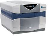
Nexcelom Celigo Micro-Well Plate Imager
 Nexcelom Celigo Image-based Microplate Reader
Function of the instrument
This instrument can do automated imaging and cell classification and counting in standard multiwell plates.
Overview
- Accepts T-flasks (T-25 and T-75) and most multi-well plates (1536-well to 6-well)
- Extremely accurate brightfield cell imaging and identification across the entire well
- Three channel fluorescence (in addition to brightfield)
- Extremely rapid scanning (~5-15 min across a range of microplates)
- Intuitive and easy-to-use, yet powerful, software segmentation and gating interface
How to Schedule
Please login to iLab system to schedule equipment time or services. For new users please follow the steps outlined in Information for New Users.
Letters of Support: We are glad to provide letters of support for grants. Please email mkirber@bu.edu. If you give us ample time we would be glad to review sections related to your imaging experiments.
Help us help you.
Grants: The Cellular Imaging Core operates at a loss and is subsidized by the Department of Medicine. What does not get included in the balance sheets are the indirect costs generated from grants obtained with the help of data from our core. You can help us continue to serve you by letting us know when you submit a proposal and/or are awarded a grant which contains data obtained from the use of our Core.
Acknowledgments: We would greatly appreciate it if authors would acknowledge the Cellular Imaging Core in their publications containing data obtained with the equipment and/or assistance of Core personnel.
Contact
Michael T. Kirber, PhD
Director of the Cellular Imaging Core (CIC)
Boston University Chobanian & Avedisian School of Medicine
Evans Biomedical Research Center – X Building
650 Albany Street
Boston MA 02118
(617) 638-7153 | mkirber@bu.edu
Location
CIC Laboratory
Evans Biomedical Research Center – X Building
650 Albany Street, Basement Room 15
Boston, MA 02118
View BUMAPS
◄ Back to Cellular Imaging Core website.
BACK TO TOP ↑