Meet the research team
The Scleroderma Research Team consists of faculty members in the Section of Rheumatology and AADC. The AADC was established in 1983 to advance basic and translational research in rheumatology and to translate laboratory findings into new therapeutic strategies for rheumatic diseases.. Their research also consists of treatment and prevention through industry-sponsored clinical trials and translational and basic research with funding support from the National Institute of Health and the National Scleroderma Foundation.
The Scleroderma Center is part of the Scleroderma Lung Study (SLS), a collaboration of 14 clinical scleroderma centers in the US whose mission is to provide SSc patients with state-of-the-art patient care and opportunities to participate in therapeutic research.
 Maria Trojanowska: Dr. Trojanowska’s research is aimed at understanding the molecular and cellular mechanisms that regulate ECM synthesis in healthy tissues and in pathological conditions such as fibrosis and tumorigenesis. The majority of her studies focus on the pathogenesis of scleroderma, an autoimmune disease characterized by vascular abnormalities and prominent fibrosis of the skin. Her laboratory uses molecular and cellular approaches and various experimental models to elucidate the mechanisms responsible for uncontrolled ECM deposition and vessel degeneration in scleroderma. The second area of investigation is related to the activation of tumour stroma. These studies examine the molecular mechanisms that mediate the controlled regulation of ECM turnover in healthy connective tissue and are responsible for the dysregulation of this process during tumorigenesis. Recent studies together with Dr. Lafyatis are examining the role of ER stress in systemic sclerosis.
 Andreea M Bujor, MD, PhD: Dr. Bujor is an Assistant Professor at Boston University Medical Center. She is a clinical rheumatologist and a physician-scientist with advanced training in scleroderma. Dr. Bujor is the supervising attending physician for the first-year rheumatology fellows during their continuity clinic and is actively engaged in the didactic core curriculum experience. In addition to teaching summer lecture series and the weekly board reviews, she also mentors fellows in scholarly activities through her basic and translational research laboratory. Her research in scleroderma myeloid dysfunction and fibrosis has been recognized as outstanding by the Rheumatology Research Foundation, receiving the Investigator Award with Malawista designation in 2020. Additionally, she received the American Heart Association Career Development Award in 2020, and the Scleroderma Clinical Trials Consortium Travel Award in 2019 with her project in scleroderma cardiomyopathy.
Established Investigator Grant (PI: Bujor) 04/2023 – 03/2025 1.80 calendar
National Scleroderma Foundation
Spatial frequency domain imaging as a new method to quantify skin changes in scleroderma
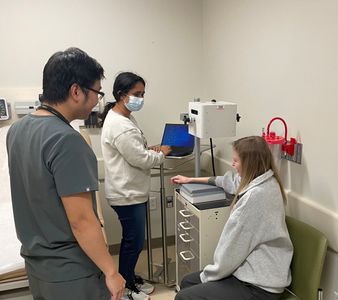
Biomedical engineering PhD student Aarohi Mehemdale and Rheumatology fellow Hung Vo using the SFDI device to measure skin changes in scleroderma patient
 Giovanni Ligresti, PhD: My laboratory focuses on lung repair and pulmonary fibrosis. We are currently investigating molecular mechanisms implicated in the differentiation of lung fibroblasts into myofibroblasts during lung fibrosis, with particular emphasis on transcriptional and epigenetic mechanisms driving persistent fibrogenesis in idiopathic pulmonary fibrosis (IPF). We have recently discovered that in the lung, myofibroblast differentiation occurs through the engagement of chromatin modifier enzymes and epigenetic readers. We found that the histone methyltransferase G9a and the epigenetic reader CBX5 play a critical role in lung fibroblast activation and that targeting these epigenetic modifiers enhanced mitochondrial function, blocked ECM protein secretion, and attenuated lung fibrogenesis.
In addition, my laboratory is interested in investigating the impact of ageing on the development of pulmonary fibrosis. We have recently discovered that the endothelium of aged mouse lungs is dysfunctional and this abnormality leads to capillary loss and persistent lung fibrosis following injury. Using a multi-omics approach (RNA-seq, ATAC-seq, and scRNA-seq) we have identified transcriptional programs in lung endothelial cells orchestrating lung vascular repair and whose abnormal activation with ageing leads to persistent lung fibrosis.
 Jeffrey Browning, PhD: My interests focus on understanding how the immune system interacts with stromal elements to form the specialized structures that orchestrate immunological encounters in lymphoid organs. On a different plane, the barriers to effective quantitation of disease are formidable in some autoimmune diseases especially those with considerable unmet need such as lupus and scleroderma. I am interested in bringing new views onto the human immune system to improve clinical studies. Mesenchymal cell differentiation pathways are intimately interwoven with pathological processes e.g. compromised vascular integrity, aberrant tissue remodeling and fibrosis and tumor-stromal interactions. In lymphoid organs, the lymphotoxin pathway, a TNF family member, is one mechanism by which both innate and adaptive lymphoid cells communicate with their stromal microenvironments. The maintenance of a differentiated follicular dendritic cell network to scaffold the B cell follicle is a well-studied example of this communication. More recently, it is clear that another network, the fibroblastoid reticular cell network, is a differentiated form of mural cells, e.g. pericytes or vascular smooth muscle cells. The precise role of the lymphotoxin pathway in controlling this structure is an area of investigation. As lymph nodes can undergo massive expansion in response to danger following by involution, they form an intriguing model of physiological tissue remodeling. Using this foundation, we have been studying the stromal underpinnings of the perivascular adventitial compartment. this compartment is considered crucial to harbor stem cells, tissue resident leucocytes and sense and initiate repair/remodelling efforts after injury. Human skin has provided an excellent tool to observe these stromal perivascular networks.