Student Updates
$4 million NIH Grant Establishes Boston Rehabilitation Outcomes Center at BUSPH
Although techniques to measure physical rehabilitation outcomes have been greatly improved in recent years, there is no gold standard to gauge the success of rehabilitation efforts for sick or injured patients.

A grant awarded to the Health & Disability Research Institute (HDR Institute) at Boston University School of Public Health aims to address this problem by establishing the Boston Rehabilitation Outcomes Center (Boston ROC), a collaborative of local institutions that will provide rehabilitation researchers with the most up-to-date outcome measurement tools.
The five-year grant in the range of $4 million is the first of its kind awarded by the National Institutes of Health's National Center for Medical Rehabilitation Research to examine rehabilitation outcome measures. Joining the HDR Institute in the work are Spaulding Rehabilitation Hospital, Harvard Medical School, and Tufts University. The award comes at a time when consumers, policy makers and insurers are pushing for more reliable ways of evaluating how well rehabilitation healthcare services serve patients.
"There's a move towards using measurement techniques that have solid scientific evidence behind them," said Director of the HDR Institute Alan Jette, PhD, MPH, professor of health policy & management, who will lead the new project. "This grant creates a resource that's not available in rehabilitation research today. Before long, we hope that people will eventually see Boston ROC as the go-to-place when it comes to information about emerging new rehabilitation outcome tools," he said.
In the last decade, there have been many advances in new techniques and methods available to assess outcomes in rehabilitation research, but many researchers continue to use older, more familiar techniques, according to Jette. There has not been a cohesive system in place to disseminate new methods and researchers are either unaware of the more recent measuring tools or aren't skilled and trained in them, he said.
Specifically, Boston ROC will pilot programs that will focus on topics such as
Evaluating new techniques (to be used in future clinical trials) to monitor the physical activity intensity of children with cerebral palsy;
Evaluating a touch screen and audio-assisted computer interface of a new outcome instrument that measures factors associated with the re-integration of Iraq and Afghanistan war veterans into their communities;
Investigating a new assessment instrument designed to evaluate the function and disability among patients undergoing hip or knee reconstruction surgery.
In addition to addressing specific outcome measurement challenges through pilot programs, Boston ROC will host visiting scientists and engage experts from participating institutions to conduct training workshops and web courses and conferences on newer measurement techniques.
"Over the years, Alan Jette and the researchers at the HDR Institute have made tremendous contributions in advancing rehabilitation science and in improving the skills of health professionals through training and by providing tools," said Robert Meenan, MD, MBA, MPH, dean of BUSPH. "This new effort, funded by a very competitive NIH grant, will fill a serious gap in the ability of researchers to measure rehabilitation outcomes precisely and effectively. Alan is a superb choice to lead this effort."
Originally a part of the Center for Rehabilitation Effectiveness at BU's College of Health & Rehabilitation Sciences: Sargent College, the Boston University HDR Institute was established in 2004 and moved its operation to BUSPH in 2006 as a part of the School's effort to increase its capacity to study disability as a public health issue. Faculty and staff at HDR Institute conduct funded research and provide education and training opportunities to students and health professionals.
Besides Jette, other BUSPH faculty involved in Boston ROC include Stephen Haley, PhD, MS, professor of health policy and management and associate director of HDR Institute; Nancy Latham, PhD, MSc, research assistant professor of health policy and management; Pengsheng Ni, MD, MPH, research assistant professor of health policy and management, and Mary Slavin, PhD, MS, research assistant professor of health policy and management.
Submitted by Elana Zak, ezak@bu.edu
3Is (Infection, Immunity, Inflammation) and Atherothrombosis: New Directions for Improving Patient Care, Sept. 7
Atherothrombosis is the main underlying cause of cardiovascular disease (CVD) and is poised to become the number one cause of death globally. The Center for Anti-Inflamatory Theraputics at the Henry M. Goldman School of Dental Medicine (GSDM) and the National Heart, Lung, and Blood Institute (NHLBI) are jointly holding a conference on 3Is (Infection, Immunity, Inflammation) and Atherothrombosis: New Directions for Improving Patient Care. The conference will take place on Tuesday, September 7, 2010, at 8:30 a.m. at the 670 Albany Street Auditorium on the Boston University Medical Campus. There is no cost for attend the conference but registration is required.
The objectives of the conference are:
Assess what is known about the role of infection, immunity, inflammation, and their interactions in the development and progression of atherothrombotic diseases;
Review emerging technologies that may help shape the field in the future;
Identify research needs related to infection, immunity, and inflammation directed at improving prevention and treatment of atherothrombotic CVD.
The conference will bring together leaders including Peter Libby, Joseph Loscalzo, Ira Tabas, Richard Cohen, Paul Ridker, Gerald Byrne, and Joseph Witztum who will address the core questions surrounding atherothrombosis at the interface of infection, immunity, and inflammation. The conference will provide conference attendees with a look beyond current state-of the-art.
Learn more about this conference at : http://dentalschool.bu.edu/research/ConferenceonInfectionImmunityInflammationandAtherothrombosis.htm
Time for a Change in US Health Care Delivery System
In a paper published in a special edition of the Journal of Public Health Dentistry released in June 2010, Professor and Chair of Health Policy & Health Services Research Dr. Raul Garcia discussed enhancements needed for oral health care services in order to eliminate oral health disparities. The article is titled, "Envisioning Success: the Future of the Oral Health Care Delivery System in the United States," and was also authored by Drs. Ronald Inge, Linda Niessen, and Dominick DePaola—all members of the Santa Fe Group, a nonprofit organization committed to serving as a catalyst for innovations in health care.

The article reviews and expands on contributions from the Institute of Medicine's 2009 workshop, "US Oral Health Workforce in the Coming Decade." At the workshop evidence was presented on the inequities in oral health outcomes in the US resulting from the inability of the current dental workforce to meet the needs of diverse, and often-times underserved, populations.
"The challenge for dentistry is to determine if innovative workforce solutions can be developed that will decrease disparities in access and improve the health of the nation as a whole," wrote Dr. Garcia. "Successful implementation of workforce innovations must include: reducing resistance to change by organized dentistry, adapting current and creating new educational and training programs, reforming policy governing dental practice, and creating a financing system that will enable the success of these changes."
The article goes on to define the metrics needed to evaluate success including: safety, lower disease incidence, lower prevalence of untreated disease, increased utilization of preventative services, decreased utilization of emergency services, enhanced access to needed services, and improved patient satisfaction and oral-health related quality of life.
The article can be found in full here.
Head Trauma Linked to ALS-Like Disease: researchers find neurological condition in former athletes
Over the past two years, BU researchers have linked head injuries in former athletes to depression and dementia, making headlines and leading to rule changes in the National Football League. Turns out that these problems may not be the only ones collision athletes like football players, boxers, and hockey players need to worry about. The researchers have now found a link between repetitive head trauma and a new form of motor neuron disease similar to amyotrophic lateral sclerosis (ALS), or Lou Gehrig’s disease. Their findings were published today in the September 2010 issue of the Journal of Neuropathology and Experimental Neurology.

“The significance of this finding is that not all ALS-like disease attacks out of the blue—sometimes it’s because of our choices in life,” says Ann McKee, a School of Medicine associate professor of neuropathology and neurology and a codirector of BU’s Center for the Study of Traumatic Encephalopathy (CSTE). “The more we know about what behaviors hasten disease, the more we can practice prevention.”
McKee and her colleagues discovered the new disease while examining the brains and spinal cords of 12 deceased athletes stored in the CSTE brain bank. Each showed evidence of the neurodegenerative disease chronic traumatic encephalopathy (CTE), once known as dementia pugilistica, or “punch-drunk syndrome.” But two former NFL players—Wally Hilgenberg of the Minnesota Vikings and Eric Scoggins, who played only three games with the San Francisco 49ers, but spent four seasons in the U.S. Football League—as well as a boxer, developed motor neuron disease late in their lives. Scoggins died in 2009, at age 49, and Hilgenberg in 2008, at 66. All three had been clinically diagnosed with Lou Gehrig’s disease, although they also showed behavioral changes and cognitive decline. Without warning, ALS attacks motor nerve cells in the brain and spinal cord, leading to muscle weakness, atrophy, and often paralysis. The mind, however, is left intact. Celebrated British physicist and author Stephen Hawking suffers from ALS.
In the three athletes diagnosed with ALS, McKee found the abnormal protein TDP-43 in the brain and spinal cord in a unique pattern and distribution, along with deposits of an abnormal form of tau protein, which can create tangles that strangle and destroy brain cells. Abnormal tau deposits, a marker of CTE, are not found in ALS. The new findings suggest that the motor neuron disease that affected the three athletes, while similar to ALS, represents a distinct disease never before described in the medical literature. McKee and her colleagues have named it chronic traumatic encephalomyelopathy (CTEM), and they believe the cause is repetitive head trauma absorbed in contact sports such as football and boxing.
Lou Gehrig’s disease, the most common form of motor neuron disease, is named after the New York Yankees legend who played in the 1920s and ’30s. Early symptoms include progressive problems with speech, swallowing, and breathing. Some 30,000 Americans, mostly men, live with the disease. The majority of cases are “sporadic,” meaning of unknown cause, although researchers believe the condition involves a complex interaction of genetic and environmental risk factors. ALS is fatal in nearly all cases.
“ALS or CTEM may be the most difficult diseases in existence to watch,” McKee says. “They are slow, agonizing deteriorations that are witnessed helplessly by loved ones.”
The risk of ALS has been reported to be higher among collision athletes and military veterans. A study of professional soccer players in Italy found that the incidence rate of ALS was 6.5 times higher than in the general population. Based on the number of retired NFL players with ALS, it’s estimated that the risk is at least eight times higher than in the adult male population, CSTE researchers say. A 2006 report by the National Academy of Sciences Institute of Medicine found that among military veterans with a history of head injuries, the risk of ALS was 2.3 times higher than normal. Since 2008, the U.S. Department of Veterans Affairs has considered ALS a presumptively compensable illness for all veterans.
“When we read reports about cases of the disease being linked to specific activities or experiences, such as the increased risk associated with military service or this one regarding professional athletes, we are reminded of just how complex of a disorder ALS is,” says Steven Perrin, CEO and chief scientific officer of the ALS Therapy Development Institute in Cambridge, Mass. “We’re hopeful that this new work may shed light on potentially exciting new possibilities for biomarker and therapy development.”
CSTE’s findings raise the question of whether Gehrig suffered from CTEM rather than the disease that bears his name. Nicknamed the “Iron Horse,” Gehrig played football at Columbia University before joining the Yankees and playing in 2,036 consecutive games, a record that stood for more than 60 years. He suffered at least five documented concussions and was reported to have been knocked unconscious for five minutes after taking a pitch to the head while not wearing a helmet. He played the next day. Gehrig died in 1941, two years after being diagnosed.
The center’s research efforts over the past two years have contributed to cultural changes in collision sports, most notably in the NFL. McKee has analyzed the brains of more than 35 athletes and found evidence of CTE in 12 of 13 former NFL players. Under mounting scientific and political pressure, the NFL changed its return-to-play rules and broadcast PSAs warning youth players of the danger of concussions last year and has been lobbying for youth concussion legislation in all 50 states. New posters spelling out the dangers of concussions and head trauma now hang in all NFL team locker rooms. In May, the league gave CSTE an unrestricted gift of $1 million for further research and has encouraged its players to donate their brains to BU after they die.
CSTE maintains a registry of more than 350 living athletes who have agreed to leave their brains and spinal cords and to participate in telephone-based interviews and assessments during life. Robert Stern, a MED associate professor of neurology and a CSTE codirector, is about to launch a study of retired NFL players; they will undergo extensive neurological, cognitive, psychiatric, cerebrospinal fluid, and neuroimaging tests.
Fellow CSTE codirector Robert Cantu, a MED clinical professor of neurosurgery, was recently appointed senior advisor to the new NFL Head, Neck and Spine Committee. Cantu, McKee, and CSTE codirector Christopher Nowinski, a former professional wrestler and president of the Sports Legacy Institute, now serve on the NFL Players Association Mackey/White Traumatic Brain Injury Committee.
Responding to the latest findings from BU, physicians Hunt Batjer and Richard Ellenbogen, cochairmen of the Head, Neck and Spine Committee, jointly commented: “We are pleased that the NFL has provided the support necessary for this research and look forward to continued discussion of the findings within our medical committee and with other researchers to better understand their clinical implications.”
The CSTE team is also vigorously researching the impacts on the nervous system of subconcussive head blows and milder brain trauma.
“If repetitive head trauma can spark this kind of neurodegeneration,” McKee says, “then by studying the effects of repetitive mild brain trauma, we can learn about the initial, early triggers of neurodegenerative disease and how to slow, reverse, and lessen them.”
This BU Today story was written by Caleb Daniloff. He can be reached at cdanilof@bu.edu.
Watch the BU Today video edited by Devin Hahn here.
Oral contraceptive use associated with increased risk of estrogen receptor negative breast cancer in African American women
Investigators from the Slone Epidemiology Center at Boston University School of Medicine (BUSM) have reported that African American women who use oral contraceptives have a greater likelihood of developing breast cancer than nonusers. The study results, recently published on-line in Cancer Epidemiology Biomarkers and Prevention, were based on data from the Black Women’s Health Study (BWHS), a large follow-up study of 59,000 African American women from across the U.S. conducted by investigators at the Slone Epidemiology Center since 1995.
The investigators followed 53,848 participants in the BWHS for 12 years, during which time 789 cases of breast cancer developed on which information on receptor status was obtained. The incidence of estrogen receptor negative cancer was 65 percent greater among women who had ever used oral contraceptives than among nonusers.
According to the BUSM researchers, the increase in risk was greatest for women who had used oral contraceptives within the previous five years and whose use had lasted 10 or more years, and the increase was greater for estrogen receptor negative than for estrogen receptor positive breast cancer. Estrogen receptor positive tumors have a better prognosis than estrogen receptor negative breast cancers.

Lead investigator Lynn Rosenberg, PhD, an associate director of the Slone Epidemiology Center and professor of epidemiology at BUSM, points out- that oral contraceptive formulations have changed over time, making it relevant to assess the effects of more recent formulations on breast cancer risk. “Some past studies found a stronger association with estrogen receptor negative breast cancer. This was the first assessment of the effect of oral contraceptive use on the incidence of breast cancer classified by receptor status among African American women,” said Rosenberg who is also the principal investigator of the BWHS. “A mechanism to explain an adverse influence of oral contraceptives on development of estrogen receptor negative breast cancer is currently unknown,” she added.
Funding for this study was provided by the National Cancer Institute.
Before Using Drugs to Prevent Heart Disease, Try Better Oral Health
In a Boston University/University of Helsinki collaboration, the oral enzyme salivary lysozyme (SLZ) was proven a stronger marker of metabolic syndrome in people with heart disease than C-reactive protein (CRP), which doctors currently look at before prescribing preventive heart disease drugs. Metabolic syndrome is a risk factor for heart disease.

“This indirectly suggests that improving oral health may be a prudent approach before dispensing expensive medicines such as statins,” says Research Associate Professor at Boston University Henry M. Goldman School of Dental Medicine (GSDM) Dr. Sok-Ja Janket “but we need to look at changes in metabolic syndrome markers with dental treatment in a clinical trial.”
SLZ was significantly associated with metabolic syndrome, even after the effect of the systemic inflammation marker CRP was controlled.
SLZ was associated with an even earlier stage of heart disease than hypertension, another risk factor for heart disease.
“In other words, SLZ is a marker for heart disease very early on,” Dr. Janket says.
Metabolic syndrome, also called Syndrome X, is characterized by three of five criteria:
- abdominal obesity
- elevated fasting glucose level or having diabetes
- high triglyceride level
- low HDL level
- high blood pressure
Dr. Janket was supported by the American Heart Association and Drs. Judith Jones, Raul Garcia, and Thomas Van Dyke co-authored the article. The first author, Dr. Qvarnstrom, is a PhD candidate at the University of Helsinki, whom Dr. Janket is mentoring.
Read an abstract of the study, “Association of Salivary Lysozyme and C-reactive Protein With Metabolic Syndrome,” here.
Tackling Child Marriage Will Take Education, Enforcement
"Cultural traditions are hard to change. We have to work within existing community structures and bring recognition to communities about how child marriage compromises opportunities and health for women and their children."

So says Anita Raj, professor of community health sciences at Boston University School of Public Health, in an article in the August 4 issue of JAMA. The article cites Raj's research in India, where almost half of women aged 20 to 24 years marry before they reach their 18th birthday, as well as related studies of the physical abuse experienced by young women in nations where child marriage is common.
It also mentions a new intervention program that Raj is involved in, which seeks to educate young men about reproductive issues. The project entails training local providers of health care -- usually men -- to counsel young husbands in rural areas about the benefits of proper health care for women, with particular attention to family planning and medical services during labor and delivery.
Violence and abuse can be diminished, Raj says in the article, only if India and other countries change their preference for giving birth to boys and make a concerted effort to allow girls to contribute to society. Ironically, many families say they want to protect their female children from sexual violence by marrying them off early.
"That motivation is entrenched in the communities, so we need to demonstrate that there are other options," she said.
The full article is available online.
Submitted by Lisa Chedekel
GSDM’s Kantarci Presents New Method for Accelerating Tooth Movement
Goldman School of Dental Medicine (GSDM) Associate Professor Dr. Alpdogan Kantarci traveled to Barcelona, Spain in mid-July to attend the International Association for Dental Research (IADR) General Session. At the meeting he co-chaired a session titled, "Biology of Tooth Movement," and also gave an oral presentation titled, "Photobiomodulation-induced Orthodontic Tooth Movement."
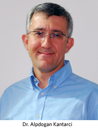
The oral presentation was based on Dr. Kantarci's research, which is sponsored by Biolux Research Ltd., and examines the effects of non-invasive photobiomodulation on tooth movement in an animal model. Photobiomodulation refers to the use of low-level, or cold, lasers that in the long-term can stimulate bone turnover, control and direct function in a pointed way, and move teeth. Dr. Kantarci found that tooth movement was significantly accelerated—a 3 to 4-fold increase—over traditional orthodontic treatment.
"The study results demonstrate the efficacy of photobiomodulation-facilitated orthodontic tooth movement at the biological level," said Dr. Kantarci. "The experimental model used in this study clearly shows that photobiomodulation increases the magnitude and rate of the tooth movement compared to the conventional technique in vivo. The findings are crucial for understanding the osteoclast-mediated bone turnover during orthodontic tooth movement, which is significantly enhanced by photobiomodulation."
Dr. Kantarci emphasized that the benefits of this type of tooth movement will be especially important for adults because they often require surgical treatment, and this non-invasive technique will provide an alternative to those who do not want to receive surgery but would like to shorten the time required for treatment.
Study Finds Acute Stress Reaction Linked to Higher Risk of Suicide
People diagnosed with acute stress reaction are more likely to complete suicide than the general population, according to a new study led by researchers affiliated with the Boston University School of Public Health.
"Clinicians and public health professionals should be aware of the importance of screening for suicidality among people immediately following a traumatic or stressful event," the authors wrote.
The study, published online ahead of print in the International Journal of Epidemiology, found that people diagnosed with acute stress reaction were 10 times more likely to complete suicide than those not diagnosed with it. The researchers also found that people diagnosed with acute stress reaction and depression or acute stress reaction and substance abuse had a greater rate of suicide.
Jaimie Gradus, DSc, MPH, an epidemiologist with VA Boston Healthcare System, assistant professor of epidemiology at BUSPH, and lead author on the study, said the paper is part of a series examining traumatic reaction diagnoses and their association with suicide. This is the first study to specifically look at the association between acute stress reaction and suicide, Gradus said.
"People who experience a trauma or have acute stress reaction seem to be at an increased risk of committing suicide, but additional research needs to be done to replicate the association," said Gradus, who is also an assistant professor of psychiatry at BU School of Medicine.
Last year, Gradus, who conducted the study while completing her doctorate in epidemiology at BUSPH, was awarded the Lilienfeld Student Paper Prize by the Society of Epidemiologic Research for her paper, "The Association between Post-Traumatic Stress Disorder and Completed Suicide in the Adult Population of Denmark."
A person is diagnosed with acute stress reaction following a traumatic experience, such as witnessing a death or being in a serious accident. Symptoms, which can include shaking, feeling that objects are unreal, irritability and problems breathing, appear within 48 hours of the event.
The authors studied 9,612 cases of suicide and 199,306 controls matched by gender, date of birth and time, taken from the nationwide Danish health and administrative registries between 1994 and 2006.
They found that 95 of the suicide cases (.99%) had acute stress reaction, compared with 165 of the controls (.08%), corresponding to a 10-times higher risk for people with acute stress reaction.
Co-authors of the study include Timothy Lash, DSc, MPH, associate professor of epidemiology at BUSPH; Henrik Toft Sørensen, MD, PhD, adjunct professor of epidemiology at BUSPH; Ping Qin, MD, PhD, MPH, associate professor of the National Centre for Register-based Research at Aarhus University in Denmark; Elizabeth Lawler, DSc, of the Department of Medicine, Harvard University; Alisa K. Lincoln, PhD, MPH, an associate professor of the health sciences and sociology at Northeastern University; Matthew Miller, ScD, MPH, MD, an associate professor of health policy and management at Harvard School of Public Health, Harvard University.
The full study is available online.
This story was submitted by Elana Zak
Director of Multimodal Whole Animal Imaging Core Appointed at Boston University’s National Emerging Infectious Diseases Laboratories
Ronald Killiany, PhD, the director of the Center for Biomedical Imaging (CBI) at Boston University Medical Campus has been appointed director of the Multimodal Whole Animal Imaging Core at the National Emerging Infectious Diseases Laboratories at Boston University (NEIDL). Killiany also serves as associate professor in the Department of Anatomy and Neurobiology at Boston University School of Medicine (BUSM) and associate professor in Environmental Health at the School of Public Health.

As director of the Multimodal Whole Animal Imaging Core, Killiany will be responsible for directing the overall whole animal imaging program including recruiting and training staff, developing services that the core will provide, writing procedural manuals and standard operating procedures for core services, and supervising experiments utilizing the whole animal imaging facilities in the NEIDL.
A joint statement from Mark Klempner, MD, Director of the NEIDL, Mark Moss, PhD, Chair of Department of Anatomy and Neurobiology, and Alex Norbash, MD, Chair, Radiology Department, said “Research on many emerging infectious diseases has been hampered by the inability to probe experimental models with modern imaging techniques. Killiany brings extensive experience with sophisticated, experimental imaging, especially MRI, to the research “toolbox” that can now be applied to these serious infectious diseases. We are excited that he will help launch the NEIDL into a leadership position for such studies.”
The primary objective of the Multimodal Whole Animal Imaging Core is to utilize newly available imaging modalities to study the pathogenesis, treatment and prevention of emerging infectious diseases. Recent and anticipated innovations in multimodal imaging technologies, including a custom-designed, one-of-a-kind BSL-4 compatible 4.7 Tesla whole animal MRI scanner, fluorescence optical tomography, and X-ray computed tomography are some of the imaging techniques that are located in the NEIDL. In-house developed multimodal imaging software packages designed specifically for emerging infectious diseases’ needs will facilitate further quantitative and multimodal analyses of data to gain insights into the pathogenesis and treatment of major infectious diseases that are public health concerns.
Killiany’s research has focused on investigating structure function relationships in the brain to uncover morphological changes that occur during aging and disease processes. He has extensive experience directing imaging studies in non-human primates and processing specimens for analysis of gross anatomy and histopathology.
Killiany received his master’s in psychology from the University of Hartford and doctorate in psychology from Northeastern University. He has held postdoctoral fellowships in neuroanatomy, neurobiology and neuropsychology at BUSM. As a research associate and professor he has published more than 60 peer-reviewed articles, contributed to numerous book chapters and served as an ad hoc reviewer on over 15 scientific journals.
*******************
The National Emerging Infectious Diseases Laboratories (NEIDL) at Boston University is part of a national network of secure facilities that will study infectious diseases – whether they occur naturally or are introduced deliberately through bioterrorism. The facility is dedicated to the development of diagnostics, vaccines and therapeutics to combat emerging and re-emerging infectious diseases. In addition to BSL-2 and BSL-3 laboratories, the NEIDL houses a BSL-4 laboratory that operates at the highest level of containment. The NEIDL adds to the growing life sciences industry in the region, throughout the Commonwealth of Massachusetts, and across the country.