Mammalian and bacterial ribosome-channel complexes
Nascent secretory proteins are translocated into the lumen of the ER in higher eukaryotes and across the plasma membrane in bacteria. Conversely, membrane proteins are gated laterally into the lipid bilayer by the Sec61 and SecYEG channels.
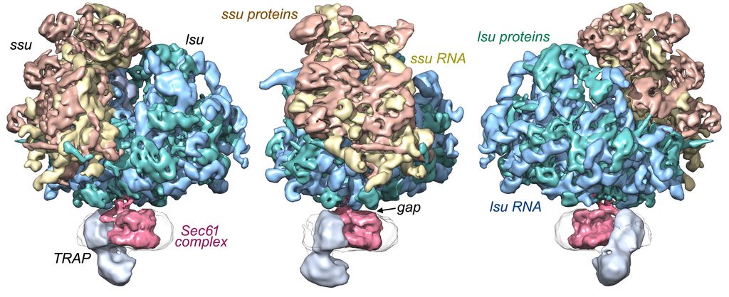
80S Mammalian ribosome channel complex
In a collaboration with Dr. Tom Rapoport (Harvard Medical School), we imaged native ribosome-Sec61 channel complexes isolated from canine endoplasmic reticulum. This allowed us to describe the architecture of the ribosome-channel complex; single Sec61 heterotrimers are present with highly conserved connections formed by the 6/7 and 8/9 loops of Sec61 alpha. A conserved gap is present between the cytoplasmic surface of the channel and the exit tunnel of the large ribosomal subunit that may allow nascent chains to access cytosolic factors during translocation. Previous work had suggested that a tight seal may be present between the large subunit and the channel. This gap may be used in quality control when large proteins are not properly folded as they enter the ER lumen and may also be used by cytoplasmic domains of membrane proteins as they begin folding during translocation. This analysis also allowed us to position single copies of Sec61 and TRAP complexes within the translocon.
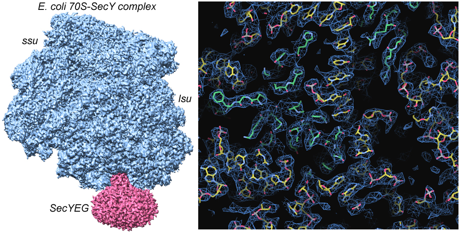
Bacterial ribosome-channel complexes
Current studies are targeting the bacterial ribosome-channel complex to provide new insights into translocation at the cytoplasmic membrane. This analysis suggests how the lateral gate of the SecYEG channel may open in the plane of the lipid bilayer.
Click on figure for animation
Selected Publications:
Park, E., Ménétret, J. F., Gumbart, J.C., Ludtke, S.J., Li, W., Whynot, A., Rapoport, T. A., and Akey, C. W. (2014). Structure of the SecY channel during initiation of protein translocation. Nature 506, 102-106. PMID: 24153188
Ménétret, J. F., Hegde, R.S., Woong, K., Gygi, S.P., Rapoport, T. A., and Akey, C. W. (2008). Single copies of Sec61 and TRAP associate with a non-translating mammalian ribosome. Structure 16, 1126-1137. PMID:18611385
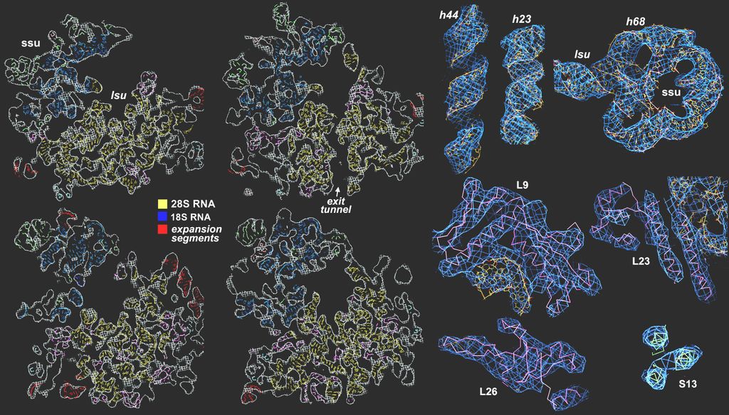
Chandramouli, P., Topf, M., Ménétret, J.F., Eswar, N., Gutell, R.R., Sali, A., and Akey, C.W. (2008). Structure of the mammalian 80S ribosome at 8.7Å resolution. Structure 16, 535-548. PMID:18400176