
Lynne M. Coluccio, Ph.D.
Associate Professor of Pharmacology, Physiology & Biophysics, Emeritus
Research
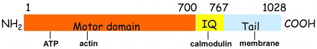
Myosins are molecular motor proteins that use the energy from ATP hydrolysis to translocate actin filaments. In general, myosins contain three domains. The head or motor domain contains the ATP- and actin-binding sites. Following the motor domain is the neck or light chain-binding domain (LCBD), which contains one or more light chain-binding or IQ motif, which binds specific light chains or calmodulin. The C-terminus consists of the tail region, which is responsible for dimerization or cargo binding.
Myosins constitute a superfamily, and dozens of different myosins have been identified. They share sequence similarity in the motor domain, although they differ in their kinetic interaction with actin. The length of the light chain-binding region and the particular light chain(s) they bind vary among myosins. In addition, depending on the myosin, the tail region can mediate dimerization or binding to various binding partners, which can be cargo, regulatory molecules, and/or lipids.
The best-known myosin is myosin-II, a two-headed molecule that self assembles into bipolar filaments. Myosin-II-containing thick filaments interact with actin-containing thin filaments to drive skeletal muscle contraction. Myosin-II isoforms also mediate smooth muscle contraction, and in non-muscle cells myosin-II isoforms mediate a number of cellular events including cytokinesis, cell migration, cell adhesion, and cell signaling.
The most diverse members of the myosin superfamily are class I myosins, which are single-headed and therefore do not form filaments like myosin-II does. The first myosin-I to be identified in mammalian cells was Myo1a, which forms the lateral links in intestinal microvilli observed between the core bindle of actin filaments and the membrane. We found some years ago that (i) expression of myosin-I is not confined to intestine as believed at that time and that (ii) cells express multiple class I myosins by discovering three different class I myosins in liver. In fact, eight class I myosins, A-E, are expressed in mammalian cells. Investigating the cellular and molecular properties of Myo1b and Myo1c has been the primary focus of our laboratory.
As part of a plan to determine their cellular roles, we examined the molecular mechanisms of Myo1b and Myo1c using purified native proteins as well as wild-type and mutant proteins expressed in the Sf9/baculovirus expression system. In transient kinetic studies done in a long-term collaboration with Michael Geeves, University of Kent, Canterbury, we showed that Myo1b and Myo1c are kinetically slow.
Because class I myosins are kinetically slow, use of these myosins in single-molecule studies done with Justin Molloy and Claudia Veigel, then at the University of York, UK, revealed for the first time that the power stroke of myosins actually consists of two separate mechanical parts, key information in understanding how myosin motors work in general.
Based primarily on the biphasic ATP-induced dissociation of Myo1b-actin and Myo1c-actin observed in kinetic studies, the two-part power stroke in single-molecule studies and cryo-EM studies performed in collaboration with Ron Milligan showing that Myo1b and Myo1c exhibit an ADP-induced conformational change, we proposed that these molecules exhibit a strain-sensitive ADP-release mechanism that allows them to adapt to mechanical load making these myosins well suited for membrane events such as maintenance of cortical tension.
The biochemical and biophysical properties of Myo1c that we determined have been used for modeling its putative role in adaptation in the hair cells of the inner ear. We characterized three Myo1c mutants that are associated with hearing loss and found that the mutations cause defects in the interaction with nucleotide and actin. From these data we predicted how the mutations may affect adaptation. Definitive evidence that Myo1c mediates adaptation awaits further investigation.
Myo1c may play a variety of roles in different cells. Myo1c was shown by the Czech group to mediate GLUT4 trafficking in adipocytes. We found that Myo1c plays a role in cell-cell contact in polarized epithelial cells. We determined that cells in which Myo1c expression is reduced by RNAi lose their polarized shape and become flattened; this effect is reversed by expression of RNAi-resistant Myo1c. Our results indicate that Myo1c stabilizes the adhesion protein E-cadherin at cell-cell contacts and therefore promotes cell polarity.
Like Myo1c, the class I myosin Myo1b also has a widespread tissue distribution. Its LCBD has 6 IQ’s; alternate splice forms contain 4 or 5 IQ domains. We found that Myo1b binds specifically and with high affinity to PIP2 and PIP3 and through mutational studies we showed that binding is via a putative PH domain in the Myo1b tail. Mutations that affect phosphoinositide binding also alter the distribution of Myo1b at the plasma membrane of HeLa cells.
We found that both the number of neutral amino acid transporters at the apical plasma membrane and the amount of amino acid transport are reduced in cultured renal proximal tubule cells expressing reduced amounts of Myo1b, evidence that Myo1b supports the association of neutral amino acid transporters with the apical plasma membrane, one of the first, if not the first, physiological role to be revealed for Myo1b.
We are currently investigating the role of Myo1b in insulin secretion. Myo1b localizes at the cell membrane and with intracellular organelles in pancreatic -cells. Myo1b depletion in cultured insulinoma (INS-1) cells dramatically reduces glucose-stimulated insulin secretion and leads to the accumulation of insulin granules in the trans-Golgi network (TGN) region of pancreatic -cells. We are currently testing the hypothesis that Myo1b may mediate the biogenesis of insulin granules or their initial transport away from the TGN.
Selected Publications
Articles:
Brady, R. J., R. H. Parsons, and L. M. Coluccio. 1981. Nocodazole inhibition of the vasopressin-induced water permeability increase of toad urinary bladder. Biochim. Biophys. Acta 646:399-401.
Koretz, J. F., L. M. Coluccio, and A. M. Bertasso. 1982. The aggregation characteristics of column-purified rabbit skeletal myosin in the presence and absence of C-protein at pH 7.0. Biophy. J. 37:433-440.
Coluccio, L. M., R. J. Brady, and R. H. Parsons. 1983. Pressure effects on the ADH-induced initiation of water flow in toad bladder. Amer. J. Physiol. (Renal, Fluid and Electrolyte Physiol.) F547-553.
Tilney, L. G., E. M. Bonder, L. M. Coluccio, and M. S. Mooseker. 1983. Actin from Thyone sperm assembles on only one end of an actin filament: a behavior regulated by profilin. J. Cell Biol. 97:112-124.
Coluccio, L. M., and L. G. Tilney. 1983. Under physiological conditions actin disassembles slowly from the nonpreferred end of an actin filament. J. Cell Biol. 97:1629-1634.
Coluccio, L. M., and L. G. Tilney. 1984. Phalloidin enhances actin assembly by preventing monomer dissociation. J. Cell Biol. 99:529-535.
Bryan, J., and L. M. Coluccio. 1985. Platelet gelsolin caps and severs actin filaments: a kinetic analysis of depolymerization. J. Cell Biol. 101:1236-1244.
Coluccio, L. M., P. Sedlar, and J. Bryan. 1986. A 45,000-mol-wt protein from unfertilized sea urchin eggs and its 1:1 actin complex differ in their action on actin filaments. J. Muscl. Res. & Cell Motil. 7:133-141.
Coluccio, L. M., and A. Bretscher. 1987. Calcium-regulated cooperative binding of the microvillar 110K-calmodulin complex to F-actin: Formation of decorated filaments. J. Cell Biol. 105: 325-334.
Krizek, J., L. M. Coluccio, and A. Bretscher. 1987. The ATPase activity of the microvillar 110K-calmodulin complex is activated by F-actin in Mg2+ and inhibited in K+-EDTA. FEBS 225:269-272.
Coluccio, L. M., and A. Bretscher. 1988. Mapping the microvillar 110K-calmodulin complex: Calmodulin-associated or -free fragments of the 110 kd polypeptide bind F-actin and retain ATPase activity. J. Cell Biol. 106:367-374.
Bullitt, E., D. DeRosier, L. M. Coluccio, and L. G. Tilney. 1988. Three-dimensional reconstruction of an actin bundle. J. Cell Biol. 107:597-611.
Coluccio, L. M., and A. Bretscher. 1989. Reassociation of microvillar core proteins: Making a microvillar core in vitro. J. Cell Biol. 108:495-502.
Coluccio, L. M., and A. Bretscher. 1990. Mapping of the microvillar 110K-calmodulin complex (brush border myosin 1). Identification of fragments containing the catalytic and F-actin-binding sites and demonstration of a calcium ion dependent conformational change. Biochemistry 29:11089-11094.
Coluccio, L. M. 1991. Identification of the microvillar 110-kDa calmodulin complex (myosin-1) in kidney. E. J. Cell Biol. 56:286-294.
Coluccio, L. M., and C. Conaty. 1993. Myosin-I in mammalian liver. Cell Motil. & Cytoskel. 24:189-199.
Williams, R., and L. M. Coluccio. 1994. Novel 130-kDa rat liver myosin-I will translocate actin filaments. Cell Motil. & Cytoskel. 27:41-48
Coluccio, L. M. 1994. Differential calmodulin binding to three myosin-I isoforms from liver. J. Cell Sci. 107:2279-2284.
Coluccio, L. M. 1994. An end in sight: Tropomodulin. (Invited mini-review) J. Cell Biol. 127:1497-1499.
Balish, M., and L. M. Coluccio. 1995. Identification of brush border myosin-I in liver and testis. Biomed. Biophys. Res. Commun. 211:331-339.
Williams, R., and L. M. Coluccio. 1995. Phosphorylation of myosin-I from rat liver by protein kinase C reduces calmodulin binding. Biochem. Biophys. Res. Commun. 216:90-102.
Coluccio, L. M. 1997. Myosin-I. (Invited review) Amer. J. Physiol. 273:C347-C359.
Veigel, C., L. M. Coluccio, J. D. Jontes, J. C. Sparrow, R. A. Milligan, and J. E. Molloy. 1999. Myosin-I produces its working stroke in two steps. Nature 398:530-533.
Coluccio, L. M., and M. A. Geeves. 1999. Transient kinetic analysis of the 130-kDa myosin I (myr 1 gene product) from rat liver: A myosin I designed for maintenance of tension? J. Biol. Chem. 274:21575-21580.
Balish, M. F., E. F. Moeller, III, and L. M. Coluccio. 1999. Overlapping distribution of the 130- and 110-kDa myosin I isoforms on rat liver membranes. Arch. Biochem. Biophys. 370:285-293.
Geeves, M. A., and L. M. Coluccio. 1999. The XXVII European Muscle Conference. (Invited conference report). J. Muscle Research and Cell Motility. 20:807-809.
Li, W., J. W. Wang, L. M. Coluccio, P. Matsudaira and R. J. Grand. 2000. Brush border myosin I: A basally localized transcript in human jejunal enterocytes. J. Histochemistry & Cytochemistry 48:89-94.
Perreault-Micale, C., A. Shushan and L. M. Coluccio. 2000. Truncation of a mammalian myosin I results in loss of Ca2+-sensitive motility. J. Biol. Chem. 275:21624-21630.
Geeves, M. A., C. Perreault-Micale, and L. M. Coluccio. 2000. Kinetic analyses of a truncated mammalian myosin I suggest a novel isomerization event preceding nucleotide binding. J. Biol. Chem. 274:21575-21580.
Wallace, M. I., C. Batters, L. M. Coluccio and J. E. Molloy. 2003. Nanometre resolution tracking of myosin-1b motility. IEE Proc. Nanobiotechnol. 150:134-140.
Batters, C., C. P. Arthur, A. Lin, J. Porter, M. A. Geeves, R. A. Milligan, J. E. Molloy, and L. M. Coluccio. 2004. Myo1c is designed for the adaptation response in the inner ear. EMBO J. 23:1433-1440.
Batters, C., M. I. Wallace, L. M. Coluccio, and J. E. Molloy. 2004b. A model of stereocilia adaptation based on single molecule mechanical studies of myosin-I. Phil. Trans. R. Soc. B. 359:1895-1905.
Stafford, W. S., M. Walker, J. Trinick and L. M. Coluccio. 2005. Mammalian class I myosin, Myo1b, is monomeric and crosslinks actin filaments as determined by hydrodynamic studies and electron microscopy. Biophys. J. 88:384-391.
Clark, R., M. Ansari, S. Dash, M. A. Geeves, and L. M. Coluccio. 2005. Loop 1 of transducer region in Myo1b modulates actin affinity, ATPase activity, and nucleotide access. J. Biol. Chem. 280:30335-30942.
Coluccio, L. M. 2006. Myo1b. AfCS-Nature Molecule Pages. (doi:10.1038/mp.a001573.01). On line at http://www.signaling-gateway.org/molecule/query?afcsid=A001573
Lieto-Trivedi, A., S. Dash and L. M. Coluccio. 2007. Myosin surface loop 4 modulates inhibition of acto-myosin 1B ATPase activity by tropomyosin. Biochemistry 46:2779-2786. PMID: 17298083
Coluccio, L. M. 2008. Myosins: A superfamily of molecular motors, Editor. Vol. 7, Proteins and cell regulation series, Springer Verlag, The Netherlands.
Adamek, N., L. M. Coluccio and M. A. Geeves. 2008. Calcium-sensitive ATP hydrolysis of Myo1c, the adaptation motor in the inner ear. Proc. Natl. Acad. Sci. (USA). 105: 5710-5715. PMID: 18391215 PMCID: PMC2299219
Lieto-Trivedi, A. and L. M. Coluccio. 2008. Calcium, nucleotide and actin affect the interaction of mammalian Myo1c with its light chain calmodulin. Biochemistry 47:10218-10226. PMID: 18729383
Adamek, N., A. Lieto-Trivedi, S. Dash, M. A. Geeves and L. M. Coluccio. 2010. Modification of loop 1 affects the nucleotide-binding properties of Myo1c, the adaptation motor in the inner ear. Biochemistry 49(5):958-71. PMID: 20039646
Wang, C.-L., A. and L. M. Coluccio. 2010. New insights into regulation of the actin cytoskeleton by tropomyosins. Int. Rev. Cell Molec. Biol. 281:91-128. Invited review. PMID:20460184
Komaba, S. and L. M. Coluccio. 2010. Localization of myosin 1b to actin protrusions requires phosphoinositide binding. J. Biol. Chem. 285:27686-27693. PMID: 20610386; PMCID: PMC2934636 [Available on 2011/9/3].
Adamek, N., M. A. Geeves and L. M. Coluccio. 2011. Myo1c mutations associated with hearing loss cause defects in the interaction with nucleotide and actin. Cell. Mol. Life Sci. 68:139-150 PMID: 20039646; PMCID: PMC2826812.
Chinthalapudi, K., M. H. Taft, R. Martin, F. K. Hartmann, S. M. Heissler, G. Tsiavaliaris, H. O. Gutzeit, H-J. Knölker, R. Fedorov, L. M. Coluccio and D. J. Manstein. 2011. Molecular mechanism of pentachloropseudilin-mediated inhibition of myosin motor activity. J. Biol. Chem. 286(34):29700-8. PMID: 21680745.
Tokuo, H. and L. M. Coluccio. 2013. Myo1c regulates E-cadherin-based cell-cell contacts in polarized MDCK cells. Mol. Biol. Cell. 24:2820-2833. PMID: 23864705; PMCID: PMC3771945
Komaba, S., and L. M. Coluccio. 2015. Myosin 1b regulates amino acid transport by associating transporters with the apical plasma membrane of kidney cells. PLoS ONE 10(9):e0138012. Doi:10.1371/journal.0138012. PMID: 26361046; PMCID: PMC4567078.
Tokuo, H., Bhawan, J., and Coluccio, L. M. 2018. Myosin-X is required for efficient melanoblast migration and melanoma initiation/metastasis. Scientific Reports 8:10449 PMID: 29993000.
Tokuo, H., Komaba, S., and Coluccio, L. M. 2021. In pancreatic -cells myosin 1b regulates glucose-stimulated insulin secretion by modulating an early step in insulin granule trafficking from the Golgi. Mol. Biol. Cell: mbcE21030094; PMID: 33826361
Chapters:
Coluccio, L. M. 2008. Myosin I. In Myosins: A superfamily of molecular motors, L. M. Coluccio, editor, Vol. 7, Proteins and cell regulation series, pp 95-124, Springer Verlag, The Netherlands.
Coluccio, L. M. 2018. Structure and function of mammalian myosins I. In Myosins: Biosynthesis, Classes and Function. David Broadbent, Editor. Nova Science Publishers, Inc. New York. p 1-88.
Coluccio, L. M. 2020. Myosins and disease. In: Myosins: A superfamily of molecular motors, Second edition. L. M. Coluccio, Editor, Advances in Experimental Medicine and Biology 1239:245-316, Springer Nature Switzerland AG.
Links:
Faculty Profile
ResearchGate
PubMed
Contact Us
Department of Pharmacology, Physiology & Biophysics
Chobanian & Avedisian School of Medicine
700 Albany Street, W410
Boston MA 02118-2518
Phone: (617) 358-8447
e-mail: coluccio@bu.edu