In vivo Electrophysiology
The Dancing Place Cells
Double click the image to see Place Cells Dance
The Laboratory of Molecular Neurobiology, under the Direction of Dr. David Farb, is exploring and advancing the use of in vivo electrophysiology as an applied technology in the preclinical drug discovery process. The major advantage of in vivo electrophysiology over measures of regional cerebral blood flow is found in the ability of this technology to differentiate the activity of inhibitory interneurons from that of excitatory pyramidal cells both across brain regions and within subregions. Preclinical animal studies using in vivo electrophysiology permit real time monitoring of neural network modulatory activity in response to systemically administered cognitive enhancers that cannot be otherwise ascertained. This exciting approach to preclinical drug discovery is both highly translational and complimentary to the results of human functional imaging studies.
In vivo electrophysiological recordings have been used to study neuronal activity and hippocampal dependent learning and memory function since the Nobel Prize winning discovery of “Place Cells” by O’Keefe and Dostrovsky in 1971. Place cells, are hippocampal pyramidal cells that fire with a greater frequency when an animal is in a specific location. These cells establish stable place fields within a given environment and change their firing patterns (“remap”) when the animal is moved to a novel location in a classic remapping paradigm such as that shown in the video below.
Remapping Paradigm Video
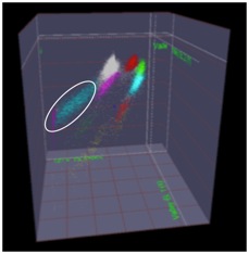
The figure below shows the place fields of a rat “remapping” when the animal is moved from a familiar square environment to a novel cylindrical one. The activity of these place cells is altered by aging and disease. Place cell activity can also be modulated with drugs (a.k.a., cognitive enhancers). In recent years, advances in vivo electrophysiology coupled with increased understanding of place cell firing dynamics has opened the door to the use of this technology as a research tool in the preclinical drug discovery process. Pyramidal cells with defined place fields in at least one environment are subsequently analyzed for drug-induced effects on firing rates, spatial correlations across environments, and spatial information content per spike.
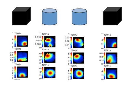
Place Fields
The Laboratory of Molecular Neurobiology acquires in vivo electrophysiological data with custom made micro-electrode arrays (such as the one pictured below). The arrays allow us to simultaneously record drug-induced changes in the activity of single place cells and local field potentials from one or more hippocampal subregions (e.g., CA3 and CA1 ) in freely behaving rodents.
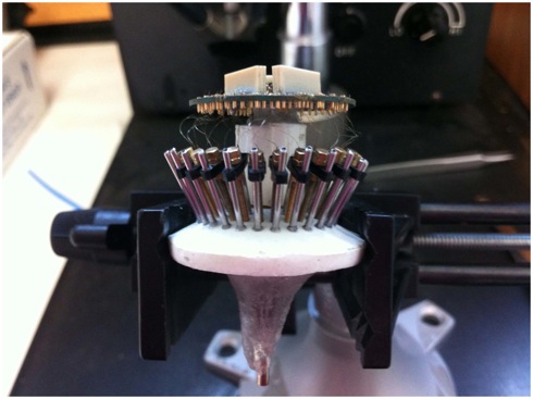
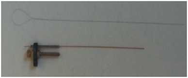
The micro-electrode arrays contain tetrodes, comprised of four nichrome micro-electrodes, such as the one pictured above (top) which can be lowered into the hippocampal subregion of interest by turning the screw on a micro drive (bottom).
Micro-electrode Array Construction Video
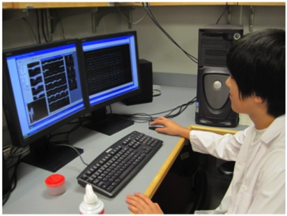
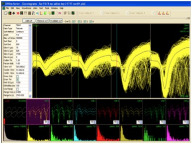
These tetrodes allow for isolation of individual pyramidal cells based on the proximity of the cell to each of the four wires of the tetrode. The action potentials or “spikes” of individual neurons such as those pictured below are “sorted” manually using Plexon Offline Sorter software as shown below or via semi-automated sorting program for subsequent analysis. Well-isolated cells such as the one pictured below show amplitude differences reflecting their proximity to the four poles of the tetrode.
Post-hoc differentiation of pyramidal “complex spike” cells (see image A below) and interneurons (see image B below) is facilitated in part by evaluation of auto-correlograms (far left panel), which show distinct patterns of activity, with pyramidal cells showing “complex spikes” characterized by bursts of action potentials immediately following the refractory period. Pyramidal cells are further differentiated from interneurons by the mean firing rates of the cell and the trough to peak width of their waveforms. Note that the pyramidal cells also have distinct place fields (far right panel) while the interneurons do not.
A.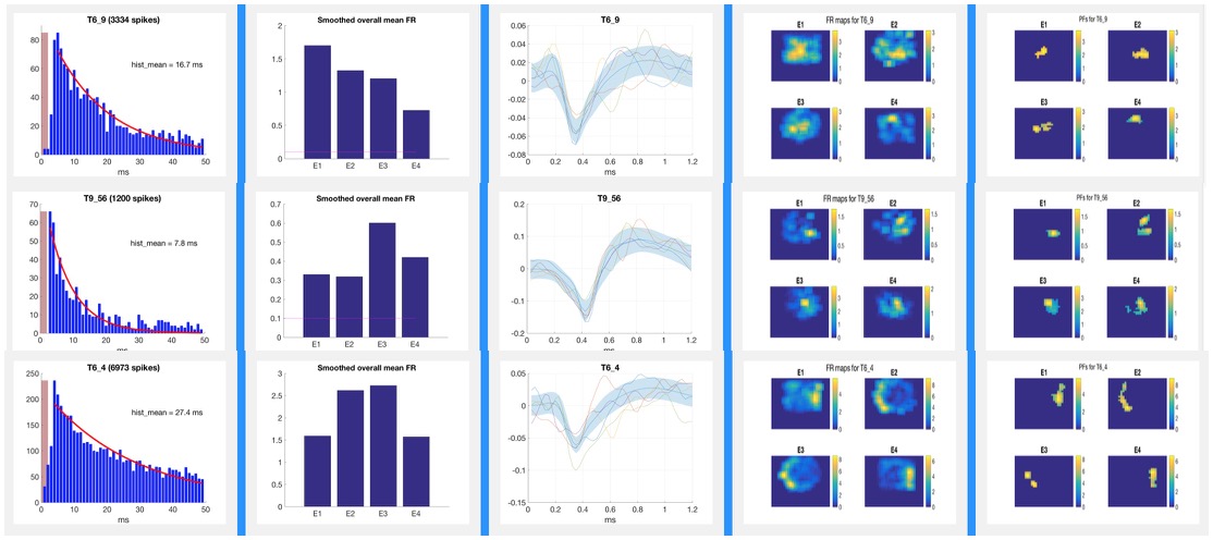
B.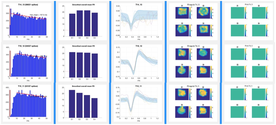
The laboratory also uses silicon probe arrays which, due to their unique design and flexible layouts permit for recording across strata within a subregion as well as from one or more subregions.
Silicon Probe Array Video
Select Publications and Abstracts
Recent News
To return to Laboratory of Molecular Neurobiology click here