Researchers Develop, Validate Tool to Visualize 3D Architectural Properties of Atherosclerosis Plaques
Atherosclerosis is a long-term arterial vessel wall disease characterized by the buildup of lipid-rich and inflamed plaques. It often goes undetected, but highly inflamed plaques disrupt and form a blood clot attached to the vessel wall adjacent to the flowing blood. This acute event (atherothrombosis) can lead to heart attack or stroke.
Boston University investigators, working with researchers from the Warren Alpert Medical School of Brown University and the Providence VA Medical Center, now have developed and substantiated an advanced magnetic resonance imaging (MRI) tool to reveal new structural insights into atherothrombosis, the leading cause of mortality in the Western world.
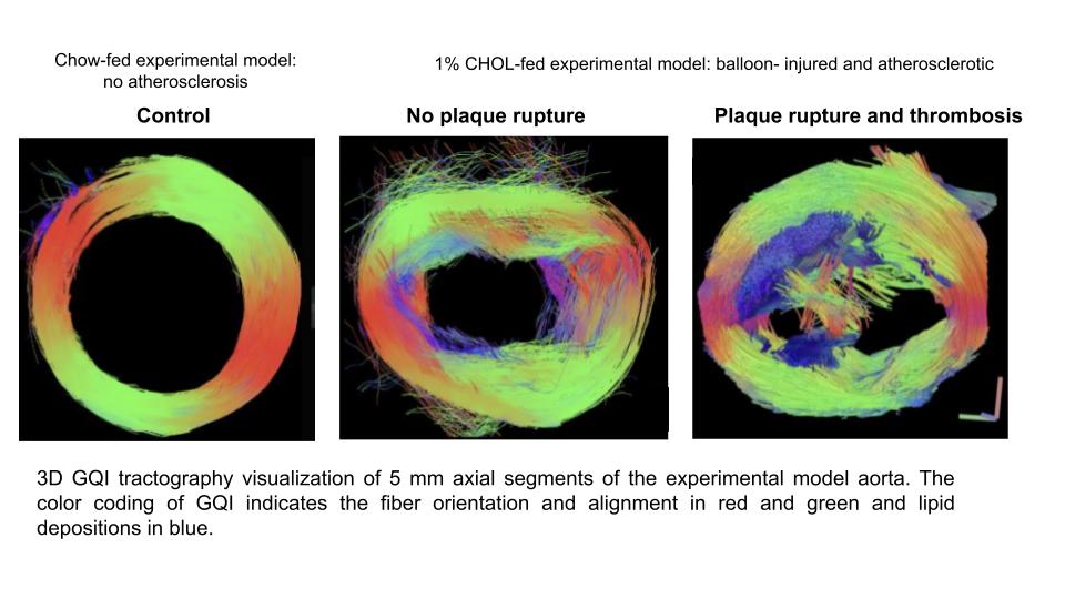
Using an experimental model, they combined magnetic resonance imaging (MRI) and mathematical analysis to architecturally define features of the fatty material that forms plaques in the arteries not visualized with conventional MRI or histology alone.
“This method uniquely detects regions of arteries at risk of rupture or atherothrombosis, thus increasing the accuracy of diagnosis and assessment of treatment outcomes in individuals with atherosclerotic disease,” explained corresponding author James A. Hamilton, PhD, professor of physiology & biophysics at Boston University Chobanian & Avedisian School of Medicine.
As atherosclerosis progresses, damaged smooth muscle cells (SMC) become inflamed and disorganized. While current bioimaging techniques focus mainly on plaque features adjacent to the flowing blood, they are unable to capture highly detailed deeper cellular elements and fibrous disorganization across the entire vessel.
In regions with normal vessel wall and low inflammation, researchers observed long-range coherence of SMC and collagen fiber orientation parallel to the vessel wall, whereas in highly inflamed regions, blood clots and underlying vessels were characterized by highly random properties with many short tracts that were perpendicular to the vessel wall.
According to the researchers, this research represents an important step forward by the Hamilton group in a decade-long collaborative project that has designed MRI methods to identify high-risk plaques that are being testing clinically.
These findings appear in Journal of Cardiovascular Magnetic Resonance