Characteristics of Amyloid Fibrils
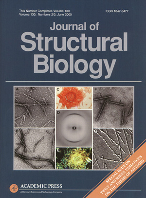
The cover of this special amyloid issue of the Journal of Structural Biology illustrates the characteristics of all amyloid fibrils. All amyloid fibrils are unbranched and are approximately 60 to 120 Å in diameter, stain with Congo red and display a green birefringence, and exhibit a cross-? structure.
Electron micrographs of (A) the amyloid fibrils of the N-terminal prion domain of a protein from yeast, (B) the amyloid fibrils of the Ab protein in Alzheimer’s disease, (F) the protofibrils of the tau protein in Alzheimer’s disease, and (G) the amyloid fibrils of amylin in type-II diabetes. The amyloid fibrils formed from synthetic Ab protein (C) stain with Congo red and (E) display a green birefringence. The amyloid fibrils formed from the de novo designed betabellin 15D exhibit a cross-? structure (D).
For more information, please visit the Journal of Structural Biology web site, or contact Cathy Costello for reprints.