Obesity-induced senescent macrophages and fibrosis
As organisms grow, older cells can undergo a phenomenon called senescence. This process defines a cell state where cells permanently stop dividing but do not die. Senescent cells secrete toxic pro-inflammatory factors contributing to the development of many diseases.
BUSM researchers have shown that obesity in experimental models led to senescence of macrophages, an immune cell subtype within fat or adipose tissue published online in the journal Life Science Alliance. Obesity-induced senescent macrophages activate a fibrotic transcriptional program in adipocyte progenitors by Nabil Rabhi, Kathleen Desevin, Anna C Belkina, Andrew Tilston-Lunel, Xaralabos Varelas, Matthew D Layne, Stephen R Farmer
According to the researchers, the fact that macrophages can become senescent is an unexpected finding. Many of the macrophages within obese tissue were senescent and those senescent cells may be a significant driver of fat tissue fibrosis. These findings suggest that obesity accelerates cellular or biological immune aging in fat.
“In healthy individuals, those cells contribute to cleaning the tissue from dead adipocytes (cells specialized for the storage of fat) and help in the cellular turnover. We demonstrated that macrophages lost this capacity when they become senescent,” explained first and co-corresponding author Nabil Rabhi, PhD, an instructor of biochemistry at BUSM.
The researchers also found that senescent macrophages secrete a variety of factors, one of which is a molecule called osteopontin which they found is responsible for adipose tissue fibrosis. “Our finding suggests that macrophages ages faster in obese animals. This accelerated senescence may contribute to the pathological thickness or fibrosis of fat tissue observed in obese individuals with type 2 diabetes,” said Rabhi.
The researchers believe understanding new regulatory pathways that control adipose tissue responses to obesity may help identify new targets for obesity treatment. “Our finding indicates that targeting the senescent macrophages population or using osteopontin inhibition may represent a promising approach for obesity treatment and its adverse complication including type 2 diabetes,” added Rabhi.
Dmitry Kretov selected for early career development award
Congratulations to Dmitry Kretov for being selected for a Department of Biochemistry Early Career Development Award. Dmitry is an Instructor working in the Cifuentes laboratory on a project entitled: “Development of an integrative approach to reveal the specificity of RNA-binding proteins and their effect on mRNA stability and translation in vivo”. This award is $21,000 for 1 year.
Nabil Rabhi selected for early career development award
Congratulations to Nabil Rabhi for being selected for a Department of Biochemistry Early Career Development Award. Nabil is an Instructor working in the Farmer laboratory on a project entitled: "Deciphering the role of lncRNAs in human adipose beige adipogenesis". This award is $21,000 for 1 year.
SARS-CoV-2 Access into Cells
The entry of severe acute respiratory syndrome coronavirus 2 (SARS-CoV-2) into human cells is an essential step for virus transmission and development of COVID 19. Although the lung epithelial cells are its initial target, SARS-CoV-2 also can infect endothelial cells. Endothelial cells are the major constituents of the vascular system and cardiovascular complication is a hallmark of severe COVID-19. Angiotensin-converting enzyme 2 (ACE2) is the entry receptor for SARS-CoV-2. However, the possible involvement of other cellular components in the viral entry is not fully understood.
A team of BUSM researchers has identified extracellular vimentin as an attachment factor that facilitates SARS-CoV-2 entry into human cells. Vimentin is a structural protein that is widely expressed in the cells of mesenchymal origin such as endothelial cells and a potential novel target against SARS-CoV-2, which could block the infection of the SARS-CoV-2.
“Severe endothelial injury, vascular thrombosis, and obstruction of alveolar capillaries (tiny air sacs scattered throughout the lungs) are common features of severe COVID-19. Identification of vimentin as a host attachment factor for SARS-CoV-2 can provide new insight into the mechanism of SARS-CoV-2 infection of the vascular system and can lead to the development of novel treatment strategies,” said co-corresponding author Nader Rahimi, PhD, associate professor of pathology & laboratory medicine.
The researchers used liquid chromatography–tandem mass spectrometry (LC-MS/MS) and identified vimentin as a protein that binds to the SARS-CoV-2 spike (S) protein and facilitates SARS-CoV-2 infection. They also found that depletion of vimentin significantly reduces SARS-CoV-2 infection of human endothelial cells. In contrast, over-expression of vimentin with ACE2 significantly increased the infection rate. “More importantly, we saw that the CR3022 antibody inhibited the binding of vimentin with CoV-2-S-protein, and neutralized SARS-CoV-2 entry into human cells,” explained Rahimi.
“The course of infection with SARS-Cov-2 is dependent on multiple factors that affect the attachment and entry of the virus into host cells, so casting a wide net and using multiple approaches to define and validate the critical components is an important strategy that can lead to the identification of protective or therapeutic targets. We show here that, in the vascular system, vimentin plays a key role in attachment of the virus to its best-known receptor, ACE2; we determined in an earlier paper in the journal ACS Central Science that CD209 and CD209L are alternative receptors that may expedite infection of tissues where ACE2 has low abundance,” said co-corresponding author Catherine E. Costello, PhD, William Fairfield Warren Distinguished Professor, Biochemistry and director of the Center for Biomedical Mass Spectrometry at BUSM.
Other collaborators from BUSM include Elke Mühlberger, PhD, and Vipul Chitalia, MD, PhD.
Story from BUSM: https://www.bumc.bu.edu/busm/2022/01/27/researchers-identify-a-new-protein-that-enables-sars-cov-2-access-into-cells/
These findings appear online in the Proceedings of the National Academy of Sciences.
Mikel Garcia-Marcos receives John Abel Award from ASPET
Congratulations to Dr. Mikel Garcia-Marcos for receiving the 2022 American Society for Pharmacology and Experimental Therapeutics (ASPET) John J. Abel Award in Pharmacology. The Abel Award is named after the founder of ASPET. It was established in 1946 to stimulate fundamental research in pharmacology and experimental therapeutics by young investigators.
The award will be presented at the ASPET Business Meeting and Awards Presentation during the ASPET Annual Meeting at Experimental Biology 2022 on Saturday, April 2 at 4:30 pm in Philadelphia. Additionally, Dr. Garcia-Marcos will deliver the Abel Award Lecture titled The Secret Life of G Proteins to open the 2022 annual meeting on Saturday, April 2 at 10:00 am in Philadelphia.
New Publication by Jarrod Moore in International Journal of Molecular Sciences
A first publication by Jarrod Moore, an MD, PhD candidate at Boston University School of Medicine and student of the Center for Network Systems Biology, in the International Journal of Molecular Sciences, looking at hypertrophic cardiomyopathy, "Mass-Spectrometry-Based Functional Proteomic and Phosphoproteomic Technologies and Their Application for Analyzing Ex Vivo and In Vitro Models of Hypertrophic Cardiomyopathy."
Hypertrophic cardiomyopathy (HCM) is an autosomal dominant disease thought to be principally caused by mutations in sarcomeric proteins. Despite extensive genetic analysis, there are no comprehensive molecular frameworks for how single mutations in contractile proteins result in the diverse assortment of cellular, phenotypic, and pathobiological cascades seen in HCM. Molecular profiling and system biology approaches are powerful tools for elucidating, quantifying, and interpreting dynamic signaling pathways and differential macromolecule expression profiles for a wide range of sample types, including cardiomyopathy. Cutting-edge approaches combine high-performance analytical instrumentation (e.g., mass spectrometry) with computational methods (e.g., bioinformatics) to study the comparative activity of biochemical pathways based on relative abundances of functionally linked proteins of interest. Cardiac research is poised to benefit enormously from the application of this toolkit to cardiac tissue models, which recapitulate key aspects of pathogenesis. In this review, we evaluate state-of-the-art mass-spectrometry-based proteomic and phosphoproteomic technologies and their application to in vitro and ex vivo models of HCM for global mapping of macromolecular alterations driving disease progression, emphasizing their potential for defining the components of basic biological systems, the fundamental mechanistic basis of HCM pathogenesis, and treating the ensuing varied clinical outcomes seen among affected patient cohorts.
Jarrod Moore is in his third doctoral year, which includes combined mass-spectrometry-based proteomics and tissue engineering training. His work is generously supported through the Kilachand Fellowship from the Multicellular Design Program at Boston University and the MD/PhD program at Boston University School of Medicine.
The article can be accessed here: https://www.mdpi.com/1422-0067/22/24/13644
Faculty Position Available
The Boston University Departments of Biology (College of Arts & Sciences) and Biochemistry (School of Medicine) invite applications for a tenure-track Assistant Professor position in Systems Biology starting in Fall 2022. We seek to recruit a colleague who uses high-throughput experimental and systems-level approaches to elucidate complex and dynamic biological processes, such as cell signaling, development in model organisms, epigenetic regulation, metabolism, nervous system function, protein homeostasis/trafficking, or transcriptional control. This search builds on recent faculty growth in systems biology and establishment of the Center for Network Systems Biology and seeks to generate new research synergies. Responsibilities include growing a vibrant research program with extramural funding, teaching graduate and/or undergraduate courses, and mentoring graduate students in research, with opportunities to participate in several interdisciplinary graduate programs. The successful candidate will be jointly appointed in Biology and Biochemistry and will contribute to a strong and growing interdisciplinary systems biology research community at Boston University that also benefits from close affiliations with engineering initiatives (including Photonics and Biological Design), and with clinical/translational efforts at the medical school. Boston University expects excellence in teaching and in research, and is committed to building a culturally, racially, and ethnically diverse scholarly community. The successful candidate will be offered newly renovated laboratory facilities as well as a competitive salary and generous start-up package.
Review of applications will begin 15 December 2021. Please use AcademicJobsOnline to submit a cover letter, curriculum vitae, statements of research, teaching interests and diversity, and three representative reprints, and arrange for three letters of reference to be submitted through the same website. In the diversity statement, applicants should provide evidence of a commitment to fostering diversity, equity, inclusive excellence, and evidence of participation in the creation of inclusive environments in their department/workplace. Inquiries can be addressed to Andrew Emili (aemili@bu.edu), Chair, Systems Biology Search Committee. Please visit the following websites for information about the Biology Department and the Department of Biochemistry.
In a continuing effort to enrich its academic environment and provide equal educational and employment opportunities, the university actively encourages applications from members of all groups underrepresented in higher education. Boston University is an AAU institution with a rich tradition of inclusion and social justice. We are proud to be the first American university to award a Ph.D. to a woman, and we continue that tradition of educating a diverse and talented student body. We are an equal opportunity employer and all qualified applicants will receive consideration for employment without regard to race, color, religion, sex, sexual orientation, gender identity, national origin, disability status, protected veteran status, or any other characteristic protected by law. We are a VEVRAA Federal Contractor.
2023 Early Career Development Awards
Last year we introduced a pilot grants program meant to enhance the careers of our postdoctoral trainees.
Again in 2023, we will award two, one-year pilot grants of $20,000 each to postdoctoral trainees for projects based on an innovative idea that establishes a new research direction. Each award will include a $1,000 supplement to the salary of the applicant. These grants are intended to provide trainees with the experience of developing a fundable research plan that can be pursued in their current labs, or included in future job applications. The guidelines of the program are as follows:
1. Eligibility: Applicants must be Postdoctoral Associates/Fellows, Instructors, or Research Assistant Professors in the BUSM Department of Biochemistry. Preference will be given to applicants who have not received previous external research funding as a Principal Investigator.
2. The project can take the applicant’s current research as a starting point, but must represent a new and innovative direction that is developed primarily by the applicant. Projects should not merely duplicate current or planned directions of the applicant’s PI.
3. Applications should include the following: (1) A research plan of no more than three pages (including figures); (2) References (no page limit); (3) A one‐page description of the applicant’s research background, accomplishments, and career plans; and (3) A budget with no more than $10,000 of salary support for the applicant. A short justification should be included for each budget item. Documents should use 11‐point font and 1⁄2 inch margins. Applicants should also include copies of their full CV.
4. A letter of support from the applicant’s PI should be included. In addition to commenting on the applicant’s qualifications, and approving the applicant’s commitment of time to the project, the letter should attest to the fact that development of the research project and preparation of the application were the work of the applicant.
Applications will be reviewed by a committee of faculty from within and outside the department, and the two selected applicants will each deliver an award lecture to the department at the end of the grant period. Feedback will be provided to all applicants by the reviewers. Interested trainees should submit a letter of intent by December 1st, including the applicant’s name, mentor’s name, and provisional title.
Full applications are due March 15th and Decisions will be announced by April 1st.
Submit Letter of Intent—Due March 1
Submit Full Application—Due March 15
Mentor—Submit Letter of Support—Due March 15
PhD Student Alejandro Rondón Ortiz Selected as Tau Leadership Fellow
“These early-career leaders in research will know their value goes beyond the bench, and that their skills and expertise can make an impact on their community and inspire future scientists” - Dr. Amy Rommel, Scientific Program Director for the Rainwater Charitable Foundation
Congratulations to Alejandro N. Rondón Ortiz, a PhD candidate in Biology-Neurobiology in both the Center for Network Systems Biology and the Laboratory of Neurodegeneration from Boston University.
The Rainwater Tau leadership fellowship is an award funded by the Rainwater Charitable Foundation. This foundation invests in early-career scientist to promote the next generation of Tau researchers/leaders. Crucial factors for the award are scientific mentorship and community outreach, which Alejandro has fulfilled by training current and future graduate and undergraduate students, especially from underrepresented identities in STEM.
Describing his research and award, Alejandro said: "For the award I used the second aim from my qualifying exam written proposal. I am using state of the art/multidisciplinary approaches to explore protein-protein interactions in neurodegeneration. I generated genetic tools that encode for chimeric proteins, and these proteins label interacting protein partners with a tag. This “tagged-protein partner” can be purified and subsequently identified by proteomics. One characteristic of these chimeric proteins is that they interact with protein aggregates and biomolecular condensates, molecular hallmarks of tauopathies. These genetic tools are predicted to work in a diversity of biological systems, including cell/neuronal cultures and brains from transgenic rodents. Finally, these uncovered pathology-associated protein networks can be cross-referenced with available datasets and propose druggable targets at early stage of the disease progression."
More about Alejandro:
He holds a PharmD degree from Universidad Catolica de Santa Maria, Peru, and an MS in Pharmacology from MCPHS University, Boston.
He is interested in neurodegenerative disorders (particularly in tauopathies). Tauopathies are a group of neurodegenerative disorders that have a common denominator: the misfolding of Tau protein (e.g. Alzheimer’s disease, progressive supranuclear palsy, cortico-basal degeneration, and others). He uses multidisciplinary approaches to explore tauopathies at the molecular level, and hopes hi findings will contribute to a better understanding of these neurodegenerative disorders.
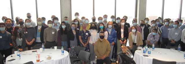
2021 Biochemistry Department Retreat
Towards the end of September the Department of Biochemistry gathered at the Science Museum in Boston for its first annual retreat in almost two years. We are a large department, and the retreat provides an opportunity to bring people together to learn about the work taking places across different laboratories and specialties, as well as meet people from the different groups. The agenda included a 'state of the department' delivered by department chair Dr. David Harris, presentations by PIs from every floor of our building, and a delicious lunch!