Nabil Rabhi awarded dKNET grant
 Congratulations to Nabil Rabhi who was recently awarded a competitive dKNET New Investigator Pilot Program in Bioinformatics Grant. This project addresses how maternal obesity impacts offspring adipose tissue cellular heterogeneity. The project will leverage the recent technological advances in single-cell and spatial transcriptomic and epigenomic to investigate how maternal metabolic status affects the composition and the function of cell types that compose adipose tissue (AT). It will uncover how maternally inherited alterations in AT cellular diversity increases progenies’ susceptibility to weight gain. CRISPR in combination with single-cell sequencing will be used to identify potential regulators of various cell states and uncover causal networks and elucidate molecular mechanisms leading to transgenerational inheritance of obesity.
New Publication – Signals promoting the development of basal-like breast cancers
A study from the Varelas Lab was recently published in Nature Communications describing the functions of the Hippo pathway effector kinases, LATS1 and LATS2 (LATS1/2), in the mammary epithelium. The study led by first author Joseph Kern tested the consequences of deleting the Lats1 and Lats2 genes in mature luminal mammary epithelia in mice. Loss of LATS1/2 led to the rapid development of basal-like carcinomas that resemble triple-negative breast cancers in humans, and over time LATS1/2-deleted tumors gained the capacity to metastasize. Luminal cells lacking LATS1/2 activity were found to exhibit luminal-to-basal plasticity, indicating that LATS1/2 are required to maintain the mature luminal mammary epithelial state. The phenotypes observed could be rescued by deletion of the transcriptional effectors YAP and TAZ, which are downstream phosphorylation targets of the LATS1/2 kinases, implicating aberrant YAP/TAZ activity in basal-like carcinoma development. Gene expression signatures derived from LATS1/2-deleted carcinomas closely resembled those of human basal-like cancers, suggesting dysregulated LATS1/2 activity is a contributing molecular event to the development of human triple-negative breast cancers.
Junior Faculty Advancement Program in Biochemistry
We announce the introduction of a new career pathway (JFA Program) for postdoctoral scientists interested in an academic career. The purpose of this program is to develop the careers of talented trainees in the Dept. of Biochemistry, with the possibility of retaining them within the Department over the long term. The JFA program is meant to provide a clearly defined pathway for academic advancement in which the trainee can progress from the postdoctoral level, through the research-track faculty level, and finally to a position as an unmodified Assistant Professor with independent space and funding.
For additional details on this program and how to apply please contact Erika Casavant, Executive Assistant to David Harris, Chair of Biochemistry erikac13@bu.edu
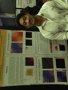
Congratulations UROP students
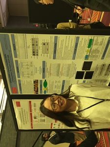
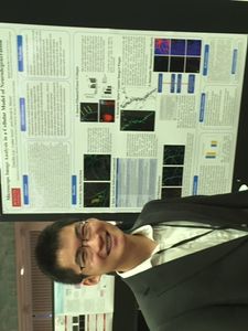
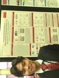
Congratulations to the UROP students that performed research in the department in the summer of 2022. Students presented their research at the annual UROP symposium on the Charles River Campus. 8 different labs hosted 12 students and their projects included: bioinformatics, mass spectrometry, G-protein signaling, and extracellular matrix biology.
Dr. Daniel Cifuentes Receives Ignition Award
Each year, BU Technology Development helps to fast-track promising new efforts by providing up to $75,000 to a few select projects. Ignition Awards provide BU faculty with these one-year grants to help launch promising new ideas into the marketplace—better positioning investigators at all stages of project development to transform their research from concept to reality. This year, Biochemistry's Dr. Daniel Cifuentes is one proud recipient. From his interview with The Brink:
Huge numbers of diseases, from neurological ailments to some forms of cancer, are caused by faulty regulation of RNA, the molecule that carries information to make new proteins in the body. If a second protein latches onto that RNA in the wrong place or at the wrong time, the information within the strand can’t be correctly read by the body, resulting in a deformed version of the protein that the RNA set out to create. In order to treat these sorts of ailments, researchers must find a specific stretch of the RNA where a drug might attach and correct the instructions it contains. Daniel Cifuentes, a MED assistant professor of biochemistry, is aiding this process by developing a new means to evaluate where and how strongly a protein attaches to RNA. Cifuentes’ new technology can decipher both the exact spot where a protein attaches to an RNA strand and how strong that attachment may be. “It used to take days to develop an assay to see if binding occurred on RNA, and even then, we didn’t know how robust that binding was,” says Cifuentes. “Our technology lets us test all of that at the same time in living cells, so researchers can more quickly develop drugs that target those spots.”
A full description of the projects can be found at The Brink.
Congratulations to Ladan Amin and Manveen Sethi on their promotion to Research Assistant Professor!
Congratulations to Ladan Amin and Manveen Sethi on their recent promotions to Research Assistant Professor!
Her main research focuses on cellular neuroscience and she is particularly interested in studying the molecular and cellular mechanism underlying neurodegenerative diseases.

Her research interest involves the identification and characterization of biomolecules such as proteins and glycans using mass spectrometry techniques and utilizing this information to understand biomolecular deregulation in disease models.
Congratulations graduates
Congratulations to the recent PhD graduates in the Biochemistry Department that received their degrees at the May 19th commencement.
Deborah Chang (Zaia lab)
Julia Bellows Hicks-Berthet (Varelas lab)
Nathan Mark Kingston (Varelas lab)
Joseph Lee Orofino (Perissi lab)
Russek Day winners 2022
The Department of Biochemistry Henry I. Russek Achievement Day Awards Committee is pleased to announce this year’s award winners:
Joseph Kern (Varelas lab) is the first prize winner
Adeline Matschulat (Varelas lab) is the second prize winner
Jarrod Moore (Emili lab) is the third prize winner.
Our congratulations go out to the winners as well as their advisors! We look forward to learning about their work on Henry I. Russek Achievement Day
Keith Tornheim named Distinguished Faculty of the Month
 The Faculty Affairs Office is pleased to announce that Associate Professor of Biochemistry Keith Tornheim, PhD, is May’s Distinguished Faculty of the Month.
Tornheim received his PhD in biochemistry from Brandeis University. He began his career at BUSM in 1981 as an assistant professor.
His nominators say, “Dr. Tornheim has contributed for 40 years to the teaching mission of Boston University School of Medicine. He currently teaches or has taught in the Medical School, Dental School, PhD, and MS programs. He has an unrivaled expertise in metabolic pathways and disease, and enzymology … and he is a fixture at our departmental and student/post-doc seminars. I am certain he has the best total attendance record of any member of our department!”
The nominators continued, “Keith is an advocate of the arts at BUSM; he is a faculty advisor of the Creative Arts Society and has both managed and contributed as a poet and sculptor to Art Days celebration for many years. Everyone on campus looks forward to this annual event. It is amazing to see the varied talents of our community and Keith’s efforts are a significant reason why these days continue.”
The 2021 Art Days theme was Antiracism, highlighting the creative BUMC community’s resolve to oppose racial hatred, bias and oppression of marginalized groups while promoting allyship, continued education and discourse. Tornheim contributed poems including Persistence of Inequality, Skin Deep, and Slave Memories.
Congratulations to Bob Varelas on his promotion to Professor of Biochemistry
Congratulations to Bob Varelas on his promotion to Full Professor of Biochemistry!
Research in the Varelas lab is focused on gaining insight into molecular mechanisms that direct cell fate decisions during animal development, homeostasis, and disease. The lab is particularly interested in understanding the functions of the transcriptional regulators YAP and TAZ (YAP/TAZ), which are key effectors of Hippo pathway signaling, and uses molecular and genetic approaches to study these factors across various aspects of biology.