Learn About In Vivo Imaging With Fluorescent and Bioluminescent Light Using IVIS
Join Anna Studwell, IVIS Core Technician/Manager on Monday, May 24, as she presents an introduction to In Vivo Imaging. This presentation covers the basic science behind bioluminescent and fluorescent imaging, a description of the imaging hardware, and an overview of analysis techniques using Living Image software. Researchers interested in tracking cancer cell metastasis and tumor growth, stem cell migration, and other research allowing fluorescent or bioluminescent labeling may find this system a useful tool for developing figures worthy of publication. A limited number of participants may sign up at the presentation for a demonstration of the instrument’s capabilities later that afternoon from 1:30-2:30 pm.
Some uses of this system include:
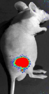
• Tracking of bioluminescent or fluorescently tagged cancer cells to monitor growth and metastasis;
• Monitoring all forms of cell transplant experiments to quantify engraftment, cell growth or cell differentiation. This includes studies of stem cell transplants, reconstitution of hematological and immune systems after irradiation.
• Monitoring the in vivo gene activity in transgenic animals carrying appropriately tagged promoter indicator trangenes.
Monitoring in vivo gene activities in cells that have been reconstituted to carry a bioluminescent or fluorescently tagged expressed gene.
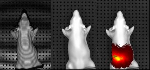
• Reconstrucing bioluminescent or fluorescent signal to a rough in vivo location in three dimensions
The presentation will take place on Monday, May 24, 2010 from 12:00 -1:00 PM, in the Wilkins Board Room, Room E120. This event is free, no registration is required. Feel free to bring your lunch.
Click here for additional information.