Plexin-A4 and Alzheimer Disease
Recently, we identified PLXNA4 as a new AD-risk gene through a genome-wide association study (GWAS) using a novel analytical approach for family-based data.1 We demonstrated that expression of this gene does not impact β-amyloid (Aβ) formation or secretion. However, the isoform encoded by the full-length transcript (TS1) when bound to its ligand SEMA3A increases phosphorylation of tau protein in neuronal cultured cells and rat hippocampal cells. In addition, TS1 expression is increased in AD compared to control brain (human post mortem study) and is correlated with tau pathology and a clinical dementia rating score (in the same subjects before death). These findings suggest that PLXNA4, and the TS1 isoform in particular, is an excellent novel target for developing a new drug that may prevent or delay onset of AD, or retard cognitive and behavioral decline.
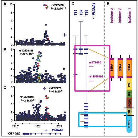
Genetic findings in the PLXNA4 region. Regional association plots of genotyped and imputed SNPs from the FS (a) and NIA-LOAD (b) datasets, and in meta-analysis (c). Most significant SNPs in the FHS (rs277470) and NIA-LOAD (rs12539196) datasets are indicated by purple diamonds. P-values are expressed as –log10(P) (y-axis) for every tested SNP ordered by chromosomal location (x-axis). Estimates of linkage disequilibrium (r2) of SNPs in this region with the top SNP computed using 1000 Genomes (hg19/Nov2010 EUR) are shown as orange circles for r2 ≥ 0.8, yellow circles for 0.5 ≤ r2 < 0.8, light blue circles for 0.2 ≤ r2 < 0.5, and blue circles for r2 < 0.2. Genomic structure of PLXNA4 was determined using the NCBI database (Build 37.1). (d) Relative position of the most significantly associated SNPs in the FS and NIA-LOAD datasets in the three validated transcripts (TS1, TS2, and TS3). Exons are denoted with horizontal bars. (e) Diagrams of functional domains encoded by amino acids from the full-length (TS1) and the shorter (TS2 and TS3) transcripts. SEMA: sema_plexinA1 interacting module (exon 1-4 in TS1 and exon 1-3 in TS2 and TS3); PSI: plexin repeat (exon 4-11 in TS1); IPT: three repeats of the binding domains of plexins and cell surface receptors (exon 11-17 n TS1); PCSR: binding domain of plexins and cell surface receptors (PCSR) and related proteins (exon 17-19 in TS1); TM: transmembrane region (exon 19 in TS1); CYTO: cytoplasmic domain (exon 20-31 in TS1).
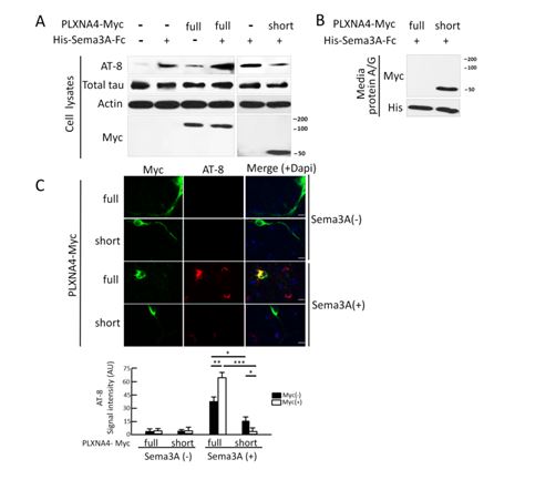
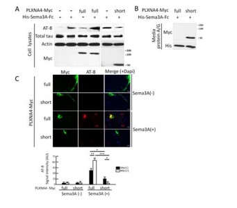 A) SH-SY5Y P301L cells were transfected with the full-length (TS1) or one of the shorter isoforms (TS2 and TS3) of PLXNA4-Myc or empty vectors (pcDNA3.1) with or without 3nM SEMA3A stimulation for 1hr. Whole cell lysates were blotted with AT8, total tau, actin and Myc. Results for the TS2 and TS3 isoforms were similar but only those for TS3 are shown. (B) 6x His-tagged Sema3A-Fc was precipitated from media by Protein A/G agarose, and the precipitates were immunoblotted with antibodies to Myc (detecting Plexin-A4 isoforms) and 6x His (detecting Sema3A-FC). (C) E18 rat primary hippocampal neurons were cultured for 14 days and transfected with the full-length or short isoforms of Plexin-A4-Myc. After transfection, cells were treated with or without 3 nM Sema3A for 1-hr, and co-stained with AT8 (1:500) and anti-Myc (rabbit polyclonal, 1:200, Covance Inc). AT8 signals were digitally captured and quantified as previously reported. Cells are immunostained with anti-Myc (green) and AT-8 (red). Scale bar represents 10 μm. *p<0.05, **p<0.01, and ***p<0.001, as determined by ANOVA and Tukey.
- Jun G, Asai H, Zeldich E, Drapeau E, Chung J, Park J-H, Chen C-D, Kim S, Haroutunian V, Foroud T, Kuwano R, Haines JL, Pericak-Vance MA, Schellenberg G, Lunetta KL, Kim J-W, Buxbaum JD, Mayeux R, Ikezu T, Abraham CR, Farrer LA. PLXNA4 is associated with Alzheimer disease risk and modulates tau protein phosphorylation. Ann Neurol 2014; 76:379-392. PMID: 25043464 http://onlinelibrary.wiley.com/doi/10.1002/ana.24219/pdf