Collaboration Encourages Museum Visitors to Feel the Science
The average person interacting with a human or animal organ could experience a range of emotions – awe, curiosity, disgust. How these feelings impact learning is the subject of a new collaboration between the Museum of Science, Boston and the Department of Anatomy and Neurobiology. Through the partnership, Museum staff are working with BUSM faculty and students to create novel displays of organ specimens and to develop informal education activities with these specimens.
Ann Zumwalt, PhD, and Jarrett Rushmore, PhD, Anatomy and Neurobiology, led a pilot of the program, which took place over the course of the past year.
“This collaboration allows us to share our anatomical expertise with museum staff, and also teaches us about public engagement with science,” said Zumwalt.
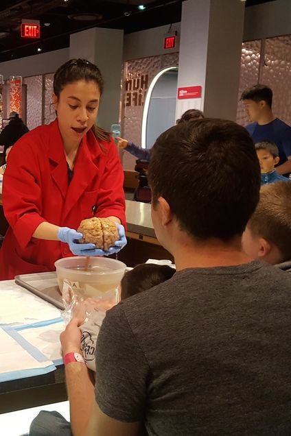
The project is centered in the Museum of Science’s Hall of Human Life, a permanent collection of more than 70 interactive exhibits that encourage visitors to explore biology and the ever-changing nature of human health and human impact on the environment. Elizabeth Kong, PhD, manager of the Hall of Human Life, leads a team of staff and volunteers who work throughout the exhibition and in the exploration hub, a popular area that features opportunities to observe and touch organs, which are part of the Museum’s collection of educational materials.
“We know that interest and experience are key to learning in informal education settings,” said Kong. “What we are currently exploring is how emotional connection can enhance learning.”
Kong and Zumwalt discuss which dissections are most desirable and appropriate for the museum environment, and BUSM faculty and students perform the dissections.
“This is a fantastic opportunity for our neuroanatomy students to perform some advanced dissections with a mind to what will be most engaging and accessible to a museum visitor,” Rushmore says.
The collaboration is currently exploring preservation techniques that will support long-term use and resilience of specimens. This aspect of the project is supported in part by a grant secured by Zumwalt from the American Association of Anatomists.
The Department of Anatomy and Neurobiology has long prioritized the training of their students to be educators in the biomedical sciences. This collaboration with the Museum has created new opportunities for students to learn about educating the museum-going public. This year BUSM graduate student Francis Zamora dissected the brains and developed activity guides for each dissection. The guides outline relevant anatomical content, discuss clinical correlations that would be interesting to the general public, and contain answers to frequently asked questions.
Anatomy & Neurobiology faculty and students will be involved in museum volunteer and staff training and will also have the opportunity to volunteer as exhibit demonstrators, giving them exposure to science outreach for the general public.
An initial pilot of the program was extremely popular, with 160 educational interactions observed in one hour. Museum staff estimates that the program has the potential to impact between 1200 and 4000 visitors each month.
Submitted by Monica Parker-James
The anatomical specimens used in this collaboration belong to the Museum and are part of their collection. They are not associated with the BUSM Anatomical Gifts program, an active anatomical donation program for the purposes of medical and dental education. Information about BUSM’s donation program may be found here: http://www.bumc.bu.edu/anatneuro/anatomical-gifts/