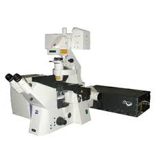
Two-Photon Confocal Microscope

This system is good for:
- Living cells
- Time lapse of fast events such as translocation, electrophysiological events
- Fixed or living samples
- Imaging at subcellular resolution
- UV excitable dyes
This system is not good for:
- Red excitable dyes
- Use of two dyes with similar emission wavelength (this system is better for single excitation dual/triple emission)
Function of Instrument
This system uses pulsed infrared light to excite dyes normally excitable in the visible and UV range. It uses a single point scanning system and multiple photomultiplier tubes to detect 3 ranges of emitted light simultaneously.
Overview
The advantages of this system are that the infrared light is well tolerated by living cells, the infrared penetrates living tissue better than visible or UV (~1mm depth), out of plane bleaching is minimized, and 3 emission wavelengths can be collected simultaneously. It is well suited for live cell and tissue imaging. The disadvantages of the system are that it is slow (several seconds per frame), it can be difficult to excite a single dye independently of others in the preparation, and it is not well suited for very low fluorescence experiments.
Training, Usage and Maintenance
- This system was developed in house and has exposed class IV laser beams. It therefore needs to be used with an operator.
- There are also sensitive photomultiplier tubes which can be destroyed with improper use.
- Regular users should have annual laser safety training.
Maintenance involves laser alignment, optical path alignment, cleaning of optics, and replacing mirrors in the light path and scanning optics. Images obtained from this instrument can be read with NIH ImageJ.
Please use the Cores Equipment Scheduler (CES) for scheduling, pricing and appointment information. For new users click here.
Help us help you.
Grants: The Cellular Imaging Core operates at a loss and is subsidized by the Department of Medicine. What does not get included in the balance sheets are the indirect costs generated from grants obtained with the help of data from our core. You can help us continue to serve you by letting us know when you submit a proposal and/or are awarded a grant which contains data obtained from the use of our Core.
Acknowledgments: We would greatly appreciate it if authors would acknowledge the Cellular Imaging Core in their publications containing data obtained with the equipment and/or assistance of Core personnel.
Location: 650 Albany Street, EBRC Building – Room 712.
Contact:
Michael T. Kirber, PhD
Core Director
Office: (617) 638-7153
Cellular Imaging Core