BUSM Neurosurgeon Removes Giant Brain Aneurysm
Imagine that over the past three years, the eyesight in your right eye has gone a little fuzzy. Your memory has dulled and you have short-term memory issues. You often blurt out inappropriate remarks. You’ve lost the mental filter that prevents faux pas. It has become challenging to perform your job. Your family thinks that your personality has changed, that you lack insight. They actually think that you have become an alcoholic! Clearly, it’s the only rational explanation for your behavior. Or, it might just be the peach-sized aneurysm in your brain.

Nirav Patel, MD, assistant professor of neurosurgery at Boston University School of Medicine, recently cared for this 55 year-old auto mechanic. A computed tomographic angiography (CTA) scan and a magnetic resonance imaging (MRI) scan of the man’s brain and vessels revealed a giant 7-cm aneurysm. Aneurysms of this size, about the dimensions of a “good-sized peach,” explains Patel, are extremely rare. “This is the largest reported aneurysm of the anterior communicating artery,” he asserts, referring to the vessel in the frontal lobes of the brain that connects two major arteries. Dysfunction in this part of the brain could readily explain why the patient was suffering both visual and cognitive difficulties.
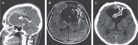
Patel performed a complex 23-hour surgery to repair the aneurysm at Boston Medical Center. The surgery greatly reduced the risk of the aneurysm bleeding or rupturing in the future, and also helped to decrease the pressure on the frontal lobe of the brain. The patient recovered extremely well from surgery and was able to return to work three months later. His vision and neurocognitive deficits improved. Repeat CT scan two years later (2015) showed complete collapse of the aneurysm sac, decreased swelling, and no evidence of new aneurysm.
In this image published in the New England Journal of Medicine on August 6, 2015, Patel explains that giant aneurysms, those greater than 2.5 cm (about 1 inch), are rare. They are associated with a high risk of rupture leading to severe disability or death if they are left untreated, so they must be repaired. As was the case with this patient, approximately 20 percent of patients with a brain aneurysm have a first-degree relative (brother, sister, mother, father, child) with an aneurysm.
Submitted by Katelyn Bird, MD.